The Coupled TuFF-BFF Algorithm for Automatic 3D Segmentation of Microglia
Publication
Metrics
AI Quick Summary
Researchers developed an automatic 3D segmentation algorithm to accurately identify microglia cells in noisy images, achieving a 20% performance increase over existing methods.
Paper Preview
Abstract
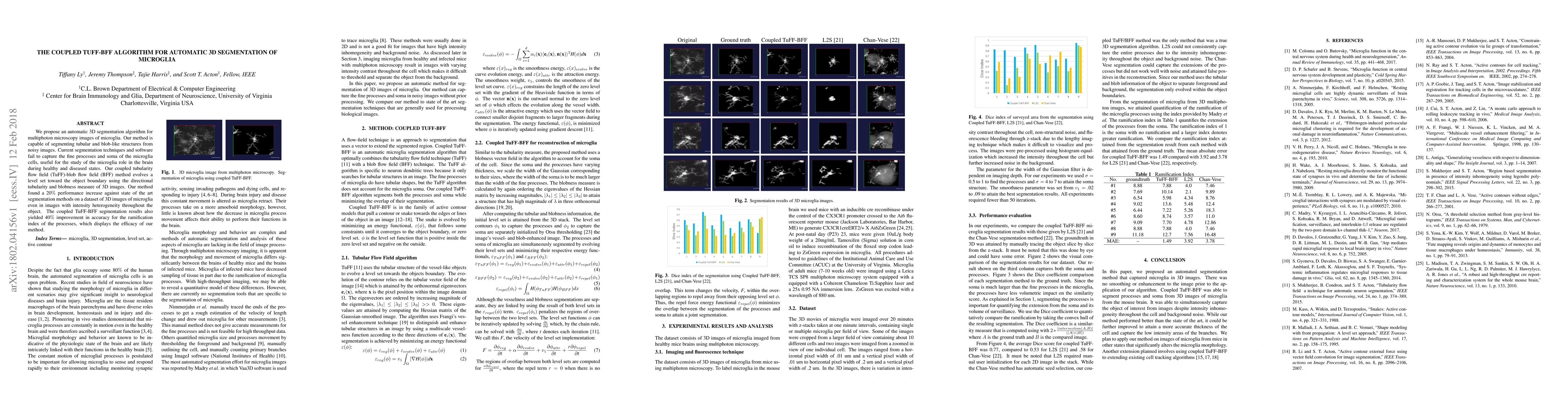
We propose an automatic 3D segmentation algorithm for multiphoton microscopy images of microglia. Our method is capable of segmenting tubular and blob-like structures from noisy images. Current segmentation techniques and software fail to capture the fine processes and soma of the microglia cells, useful for the study of the microglia role in the brain during healthy and diseased states. Our coupled tubularity flow field (TuFF)-blob flow field (BFF) method evolves a level set toward the object boundary using the directional tubularity and blobness measure of 3D images. Our method found a 20% performance increase against state of the art segmentation methods on a dataset of 3D images of microglia even in images with intensity heterogeneity throughout the object. The coupled TuFF-BFF segmentation results also yielded 40% improvement in accuracy for the ramification index of the processes, which displays the efficacy of our method.
AI Key Findings
Get AI-generated insights about this paper's methodology, results, significance, and more — seven facets brought into focus.
Impact
Paper Details
PDF Preview
Key Terms
Citation Network
Current paper (gray), citations (green), references (blue)
Display is limited for performance on very large graphs.
Discussion 0