Publication
Metrics
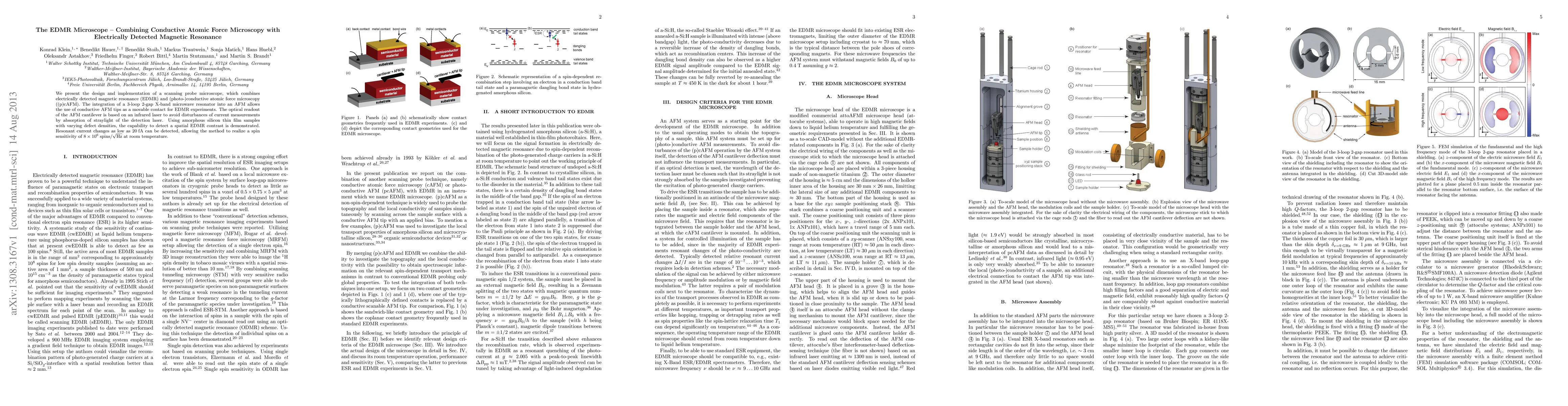
Paper Preview
Abstract
We present the design and implementation of a scanning probe microscope, which combines electrically detected magnetic resonance (EDMR) and (photo-)conductive atomic force microscopy ((p)cAFM). The integration of a 3-loop 2-gap X-band microwave resonator into an AFM allows the use of conductive AFM tips as a movable contact for EDMR experiments. The optical readout of the AFM cantilever is based on an infrared laser to avoid disturbances of current measurements by absorption of straylight of the detection laser. Using amorphous silicon thin film samples with varying defect densities, the capability to detect a spatial EDMR contrast is demonstrated. Resonant current changes as low as 20 fA can be detected, allowing the method to realize a spin sensitivity of 8*10^6 spins/Hz^0.5 at room temperature.
AI Key Findings
Get AI-generated insights about this paper's methodology, results, significance, and more — seven facets brought into focus.
Impact
Paper Details
PDF Preview
Key Terms
Citation Network
Current paper (gray), citations (green), references (blue)
Display is limited for performance on very large graphs.
Discussion 0