ArxivLens

Quick Summary:

The SLcam is a full-field energy dispersive X-ray camera that enables real-time visualization of elemental distribution in samples through fast, full-field X-ray fluorescence imaging without scanning, offering high spatial resolution and a modular setup.

DOI: 10.1088/1748-0221/7/11/C11008
arXiv Id: 1210.4344
Comments: 4 pages, 2 figures, iWorid 2012 confernce contribution
Date Published: 2015-06-11
Date Updated: 2015-06-11

Key Terms (TF-IDF):

optics (0.274)
camera (0.265)
ray (0.222)
imaging (0.178)
resolution (0.155)
dispersive (0.149)
sample (0.148)
fluorescence (0.146)
single photon (0.142)
distinguished (0.142)
space (0.140)
dedicated (0.139)
scanning (0.135)
window (0.132)
shaped (0.131)
acquisition (0.131)
course (0.129)
setup (0.126)
modular (0.125)
laboratory (0.125)
License: http://arxiv.org/licenses/nonexclusive-distrib/1.0/

Summary:

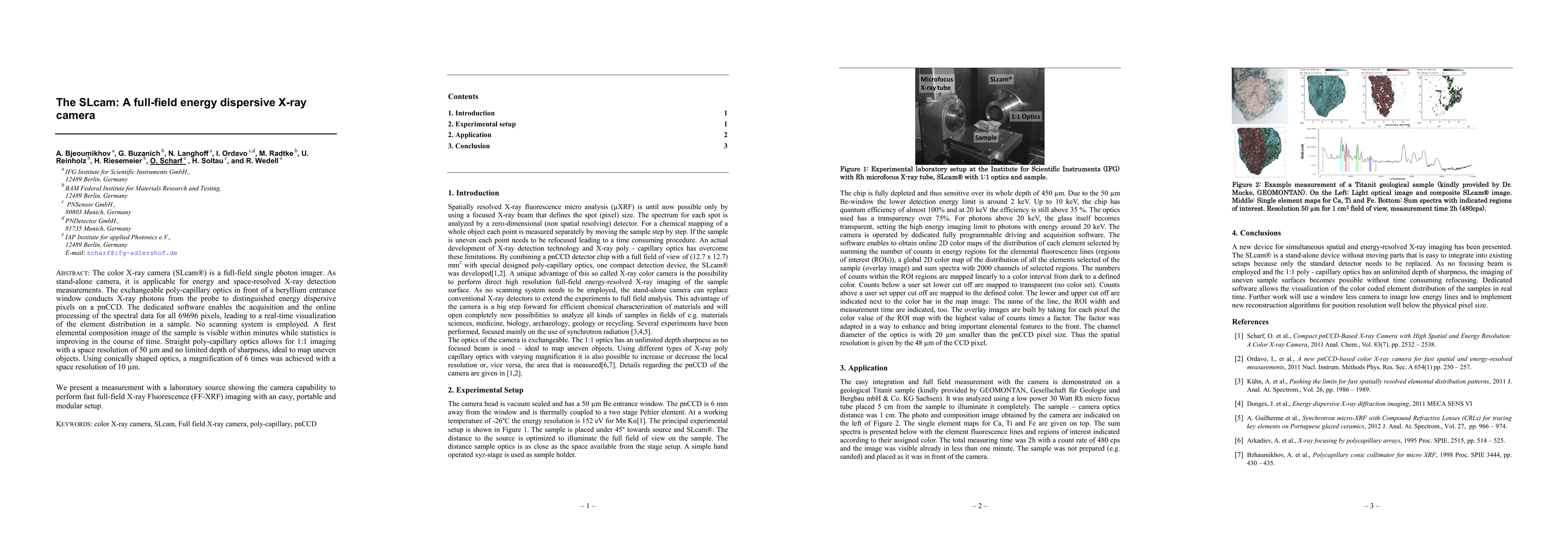
The color X-ray camera (SLcam) is a full-field single photon imager. As stand-alone camera, it is applicable for energy and space-resolved X-ray detection measurements. The exchangeable poly-capillary optics in front of a beryllium entrance window conducts X-ray photons from the probe to distinguished energy dispersive pixels on a pnCCD. The dedicated software enables the acquisition and the online processing of the spectral data for all 69696 pixels, leading to a real-time visualization of the element distribution in a sample. No scanning system is employed. A first elemental composition image of the sample is visible within minutes while statistics is improving in the course of time. Straight poly-capillary optics allows for 1:1 imaging with a space resolution of 50 um and no limited depth of sharpness, ideal to map uneven objects. Using conically shaped optics, a magnification of 6 times was achieved with a space resolution of 10 um. We present a measurement with a laboratory source showing the camera capability to perform fast full-field X-ray Fluorescence (FF-XRF) imaging with an easy, portable and modular setup.

Citation & Reference Network Visualization

This graph shows how this paper is connected to other research. The current paper is shown in dark gray, papers that cite it are in green, and papers it references are in blue. You can:

Current Paper
Papers citing this work (Citations)
Papers cited by this work (References)

• Hover over any node to see the paper title

• Click a node to view that paper's details

• Drag nodes to rearrange the graph

• Use the zoom controls (bottom right) to zoom in/out

Citations and References

Title Authors Year Actions
Spectral X-ray Detectors Based on Multi-Element Chromium Compensated Gallium Arsenide Sensors and Application Specific Integrated Circuits: : Keynote Paper Devoted 75th Anniversary of the Transistor
L. Shaimerdenova, A. Vinnik, M. Skakunov, O. Tolbanov, A. Tyazhev, Andrey V. Zarubin
2022
View Paper
Color ptychography with a hyperspectral x-ray camera
Wiebe Stolp, S. Cipiccia, S. Vanheule, D. Batey, F. Van Assche, M. Boone
2021
View Paper
X-Ray Ptychography with a Laboratory Source.
D. Batey, F. Van Assche, S. Vanheule, M. Boone, A. Parnell, O. Mykhaylyk, C. Rau, S. Cipiccia
2021
View Paper
Full-field X-ray fluorescence imaging with a straight polycapillary X-ray collimator
S. An, D. Krapohl, B. Norlin, G. Thungström
2020
View Paper
Full field x-ray fluorescence imaging using micro pore optics for planetary surface exploration
P. Sarrazin, D. Blake, M. Gailhanou, P. Walter, E. Schyns, F. Marchis, K. Thompson, T. Bristow
2017
View Paper
Röntgenabsorptionsspektroskopie im Labor
C. Schlesiger
2019
View Paper
Spectroscopic and Microscopic X-ray Fluorescence Analysis for Environ- mental and Industrial Applications
Siwen An, D. Krapohl
View Paper

Comments (0)

This website uses cookies to ensure you get the best experience. Learn more