Publication
Metrics
AI Quick Summary
This paper introduces a novel clustering method for time-domain feature extraction in Photoacoustic Remote Sensing Microscopy, enabling the identification of biomolecule characteristics from nanosecond-scale optical perturbations. The method allows for virtual tissue labeling and colorized visualizations that distinctly highlight different tissue components in murine brain tissue.
Paper Preview
Abstract
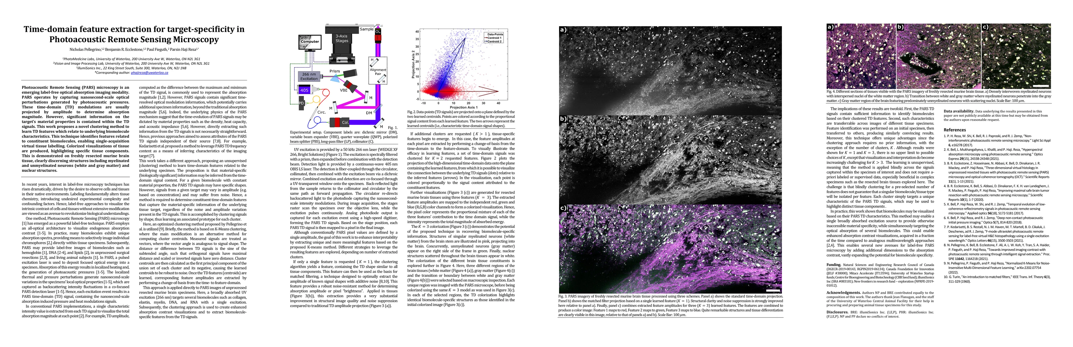
Photoacoustic Remote Sensing (PARS) microscopy is an emerging label-free optical absorption imaging modality. PARS operates by capturing nanosecond-scale optical perturbations generated by photoacoustic pressures. These time-domain (TD) modulations are usually projected by amplitude to determine absorption magnitude. However, significant information on the target's material properties is contained within the TD signals. This work proposes a novel clustering method to learn TD features which relate to underlying biomolecule characteristics. This technique identifies features related to constituent biomolecules, enabling single-acquisition virtual tissue labelling. Colorized visualizations of tissue are produced, highlighting specific tissue components. This is demonstrated on freshly resected murine brain tissue, clearly discerning structures including myelinated and unmyelinated neurons (white and gray matter) and nuclear structures.
AI Key Findings
Get AI-generated insights about this paper's methodology, results, significance, and more — seven facets brought into focus.
Impact
Paper Details
Authors
PDF Preview
Key Terms
Citation Network
Current paper (gray), citations (green), references (blue)
Display is limited for performance on very large graphs.
Discussion 0