Publication
Metrics
Paper Preview
Abstract
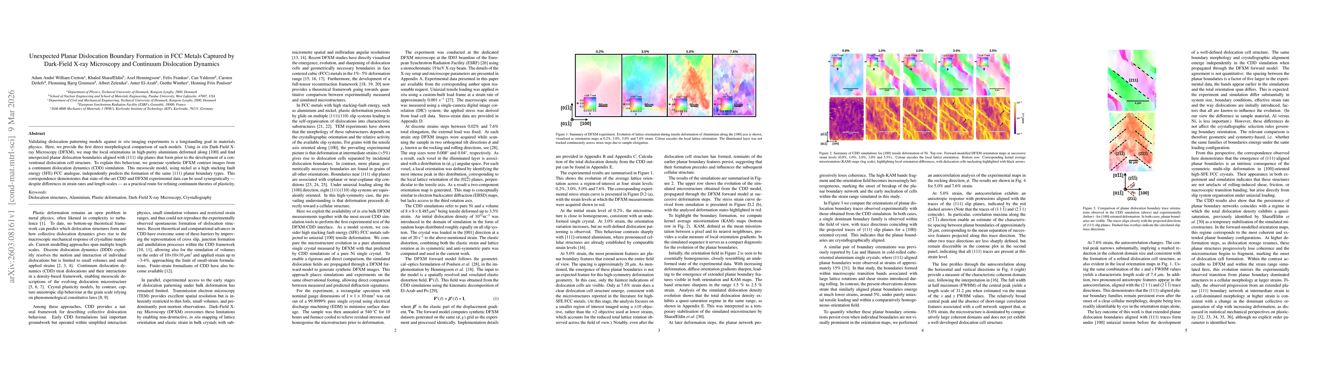
Validating dislocation patterning models against in situ imaging experiments is a longstanding goal in materials physics. Here, we provide the first direct morphological comparison of such models. Using in situ Dark-Field X-ray Microscopy (DFXM), we map the local orientations in high-purity aluminium deformed along [100] and find unexpected planar dislocation boundaries aligned with {111} slip planes that form prior to the development of a conventional dislocation cell structure. To explain this behaviour, we generate synthetic DFXM contrast images from a continuum dislocation dynamics (CDD) simulation. This mesoscale model, using nickel as a high stacking fault energy (SFE) FCC analogue, independently predicts the formation of the same {111} planar boundary types. This correspondence demonstrates that state-of-the-art CDD and DFXM experimental data can be used synergistically - despite differences in strain rates and length scales - as a practical route for refining continuum theories of plasticity.
AI Key Findings
Get AI-generated insights about this paper's methodology, results, significance, and more — seven facets brought into focus.
Discussion 0