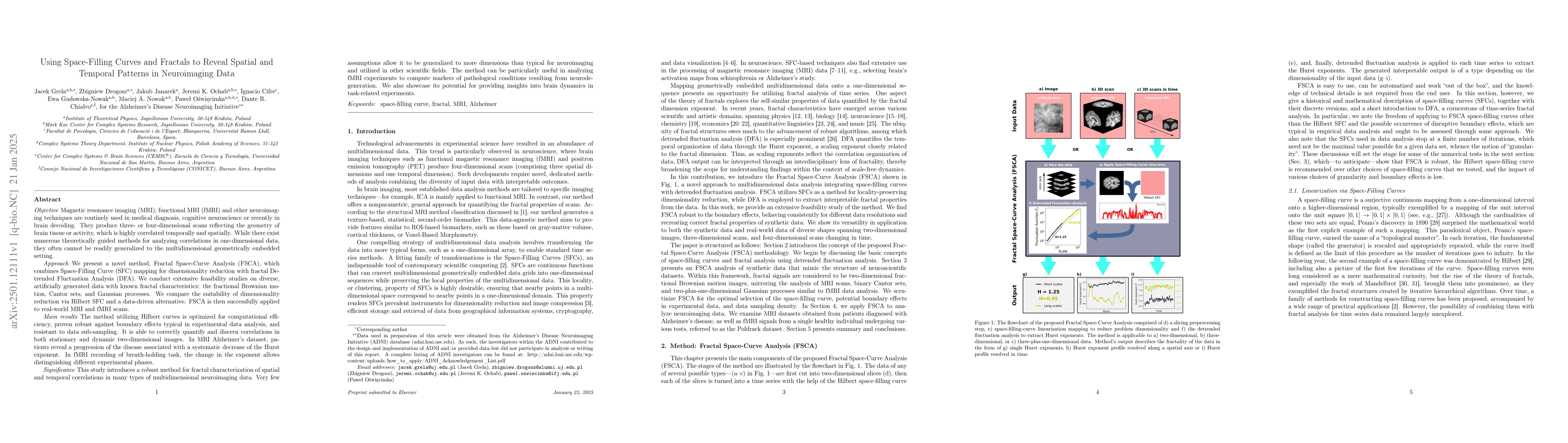
We present a novel method, Fractal Space-Curve Analysis (FSCA), which
combines Space-Filling Curve (SFC) mapping for dimensionality reduction with
fractal Detrended Fluctuation Analysis (DFA). The method is suitable for
multidimensional geometrically embedded data, especially for neuroimaging data
which is highly correlated temporally and spatially. We conduct extensive
feasibility studies on diverse, artificially generated data with known fractal
characteristics: the fractional Brownian motion, Cantor sets, and Gaussian
processes. We compare the suitability of dimensionality reduction via Hilbert
SFC and a data-driven alternative. FSCA is then successfully applied to
real-world magnetic resonance imaging (MRI) and functional MRI (fMRI) scans.
The method utilizing Hilbert curves is optimized for computational
efficiency, proven robust against boundary effects typical in experimental data
analysis, and resistant to data sub-sampling. It is able to correctly quantify
and discern correlations in both stationary and dynamic two-dimensional images.
In MRI Alzheimer's dataset, patients reveal a progression of the disease
associated with a systematic decrease of the Hurst exponent. In fMRI recording
of breath-holding task, the change in the exponent allows distinguishing
different experimental phases.
This study introduces a robust method for fractal characterization of spatial
and temporal correlations in many types of multidimensional neuroimaging data.
Very few assumptions allow it to be generalized to more dimensions than typical
for neuroimaging and utilized in other scientific fields. The method can be
particularly useful in analyzing fMRI experiments to compute markers of
pathological conditions resulting from neurodegeneration. We also showcase its
potential for providing insights into brain dynamics in task-related
experiments.
Discussion 0