Visualization of Ion Channels in Membranes using Electrochemical Strain Microscopy
Publication
Metrics
AI Quick Summary
This paper utilizes electrochemical strain microscopy (ESM) and conductive atomic force microscopy (C-AFM) to visualize hydrophilic ion channels in Nafion-phosphotungstic acid composite electrolytes. The findings reveal fibrillar ion channels formed by interactions between PWA and Nafion's sulfonic groups, enhancing understanding of ionic conduction in polymer electrolytes.
Paper Preview
Abstract
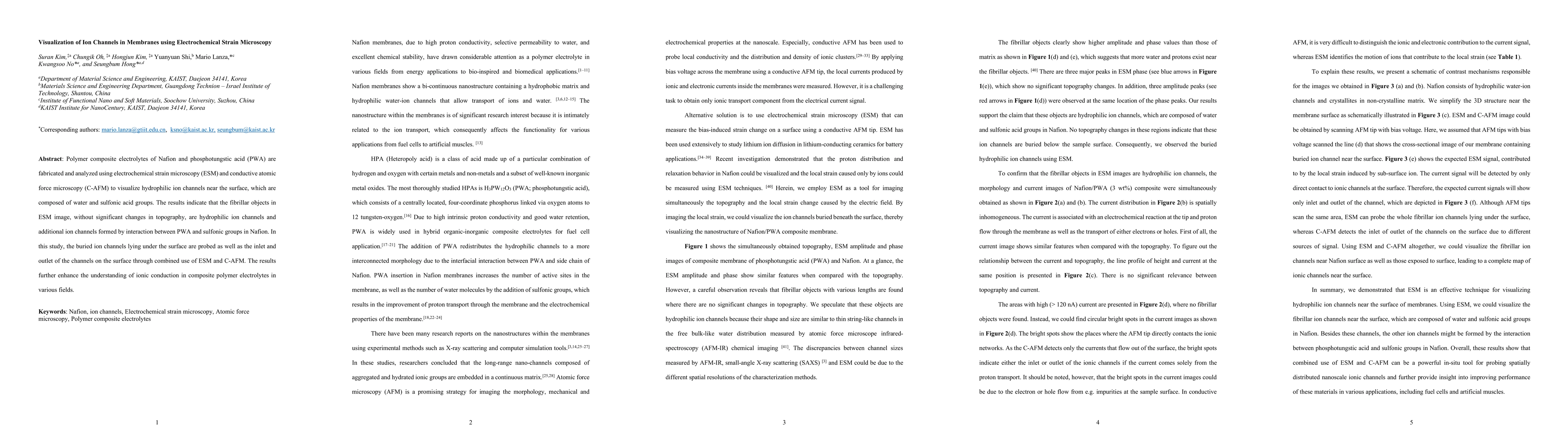
Polymer composite electrolytes of Nafion and phosphotungstic acid (PWA) are fabricated and analyzed using electrochemical strain microscopy (ESM) and conductive atomic force microscopy (C-AFM) to visualize hydrophilic ion channels near the surface, which are composed of water and sulfonic acid groups. The results indicate that the fibrillar objects in ESM image, without significant changes in topography, are hydrophilic ion channels and additional ion channels formed by interaction between PWA and sulfonic groups in Nafion. In this study, the buried ion channels lying under the surface are probed as well as the inlet and outlet of the channels on the surface through combined use of ESM and C-AFM. The results further enhance the understanding of ionic conduction in composite polymer electrolytes in various fields.
AI Key Findings
Get AI-generated insights about this paper's methodology, results, significance, and more — seven facets brought into focus.
Impact
Paper Details
PDF Preview
Key Terms
Citation Network
Current paper (gray), citations (green), references (blue)
Display is limited for performance on very large graphs.
Discussion 0