Visualizing Encapsulated Graphene, its Defects and its Charge Environment by Sub-Micrometer Resolution Electrical Imaging
Publication
Metrics
AI Quick Summary
This paper presents a method combining Kelvin force probe microscopy (KPFM) and electrostatic force microscopy (EFM) to visualize encapsulated graphene with sub-micrometer resolution, identifying defects and charge environments, crucial for high-quality device fabrication. The technique effectively detects structural defects and contaminants within the graphene flakes.
Paper Preview
Abstract
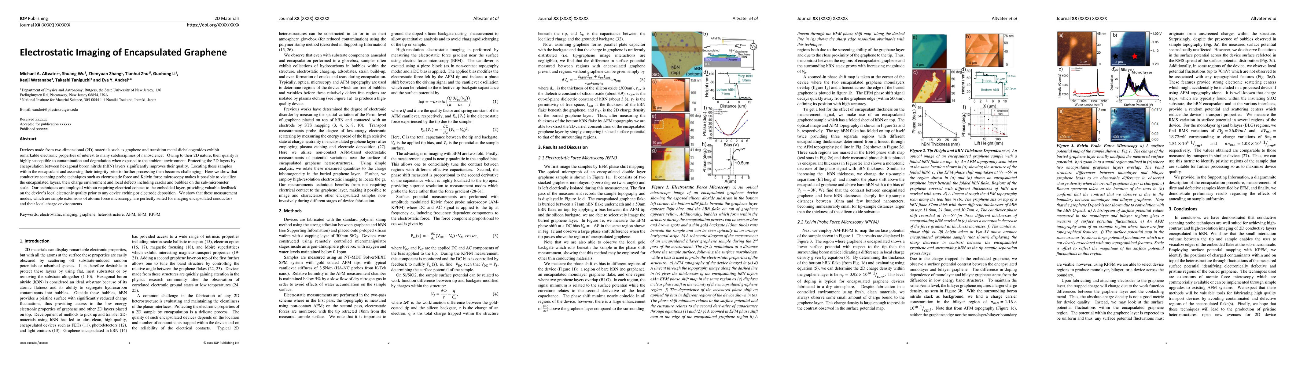
Devices made from two-dimensional (2D) materials such as graphene or transition metal dichalcogenides possess interesting electronic properties that can become accessible to experimental probes when the samples are protected from deleterious environmental effects by encapsulating them between hexagonal boron nitride (hBN) layers. While the encapsulated flakes can be detected through post-processing of optical images or confocal Raman mapping, these techniques lack the sub-micrometer scale resolution to identify tears, structural defects or impurities, which is crucial for the fabrication of high-quality devices. Here we demonstrate a simple method to visualize such buried flakes with sub-micrometer resolution, by combining Kelvin force probe microscopy (KPFM) with electrostatic force microscopy (EFM). KPFM, which measures surface potential fluctuations, is extremely effective in spotting charged contaminants within and on top of the heterostructure, making it possible to distinguish contaminated regions in the buried flake. When applying a tip bias larger than the surface potential fluctuations, EFM becomes extremely efficient in highlighting encapsulated flakes and their sub-micron structural defects. We show that these imaging modes, which are standard extensions of atomic force microscopy (AFM), are perfectly suited for locating encapsulated conductors, for visualizing nanometer scale defects and bubbles, and for characterizing their local charge environment.
AI Key Findings
Get AI-generated insights about this paper's methodology, results, significance, and more — seven facets brought into focus.
Impact
Paper Details
PDF Preview
Key Terms
Citation Network
Current paper (gray), citations (green), references (blue)
Display is limited for performance on very large graphs.
Discussion 0