This paper presents a method to visualize individual microtubules using conventional bright-field microscopy, enhanced by digital image processing to remove background noise and improve contrast. The technique is cost-effective and suitable for student laboratories, demonstrating its application in tracking microtubule motions driven by kinesin.
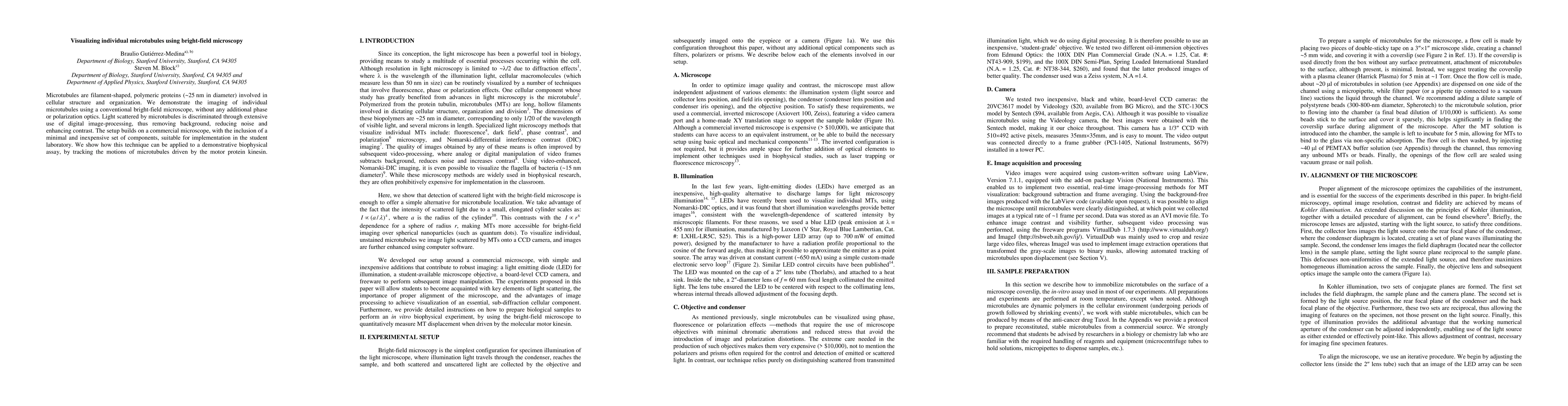
Microtubules are filament-shaped, polymeric proteins (~25 nm in diameter) involved in cellular structure and organization. We demonstrate the imaging of individual microtubules using a conventional bright-field microscope, without any additional phase or polarization optics. Light scattered by microtubules is discriminated through extensive use of digital image-processing, thus removing background, reducing noise and enhancing contrast. The setup builds on a commercial microscope, with the inclusion of a minimal and inexpensive set of components, suitable for implementation in the student laboratory. We show how this technique can be applied to a demonstrative biophysical assay, by tracking the motions of microtubules driven by the motor protein kinesin.
Get AI-generated insights about this paper's methodology, results, and significance.
Current paper (gray), citations (green), references (blue)
Amer Al-Hiyasat, Yazgan Tuna, Jonathon Howard
Gerard Glowacki, Chloe He, Alexis Gkantiragas
Title | Authors | Year | Actions |
---|---|---|---|
Approaches to visualize microtubule dynamics in vitro
|
M. Anisimov
|
2022
|
View Paper |
Kinesin-propelled label-free microtubules imaged with interference reflection microscopy
|
Gadiel Saper, H. Hess
|
2020
|
View Paper |
Quantifying bio-filament morphology below the diffraction limit of an optical microscope using out-of-focus images.
|
Madeleine Anthonisen, Yuning Zhang, M. Hussain Sangji, P. Grütter
|
2020
|
View Paper |
Interferometric Scattering (iSCAT) Microscopy and Related Techniques
|
Richard W. Taylor, V. Sandoghdar
|
2018
|
View Paper |
Optical tweezers combined with interference reflection microscopy for quantitative trapping and 3D imaging
|
Steve Simmert
|
2018
|
View Paper |
LED-based interference-reflection microscopy combined with optical tweezers for quantitative three-dimensional microtubule imaging.
|
Steve Simmert, M. K. Abdosamadi, G. Hermsdorf, E. Schäffer
|
2018
|
View Paper |
LED-based interference-reflection microscopy combined with optical tweezers for quantitative three-dimensional single microtubule imaging
|
Steve Simmert, M. K. Abdosamadi, G. Hermsdorf, E. Schäffer
|
2018
|
View Paper |
Label-free Imaging and Bending Analysis of Microtubules by ROCS Microscopy and Optical Trapping.
|
Matthias D. Koch, A. Rohrbach
|
2018
|
View Paper |
Native kinesin‐1 does not bind preferentially to GTP‐tubulin‐rich microtubules in vitro
|
Qiaochu Li, S. J. King, Jing Xu
|
2017
|
View Paper |
Transport of Intensity Equation Microscopy for Dynamic Microtubules
|
Q. Tyrell Davis
|
2017
|
View Paper |
Label-free Imaging of Microtubules with Sub-nm Precision Using Interferometric Scattering Microscopy.
|
J. Andrecka, J. Ortega Arroyo, Katie Lewis, R. Cross, P. Kukura
|
2016
|
View Paper |
Constructing 3D microtubule networks using holographic optical trapping
|
J. Bergman, O. Osunbayo, M. Vershinin
|
2015
|
View Paper |
Preparation of segmented microtubules to study motions driven by the disassembling microtubule ends.
|
Vladimir A Volkov, Anatoly V. Zaytsev, E. Grishchuk
|
2014
|
View Paper |
Imaging molecular motor regulation at the single molecule level
|
J. H. Walther
|
2013
|
View Paper |
A Minimal Optical Trapping and Imaging Microscopy System
|
Carmen Noemí Hernández Candia, Sara Tafoya Martínez, B. Gutiérrez-Medina
|
2013
|
View Paper |
Bright-Field Microscopy
|
R. Wayne
|
2019
|
View Paper |
Image Processing and Analysis
|
Randy Wayne
|
2019
|
View Paper |
Label-free imaging of microtubules with subnanometer precision using interferometric scattering microscopy
|
J. Andrecka, J. O. Arroyo, Katie Lewis, R. Cross, Philipp, Kukura
|
2016
|
View Paper |
Comments (0)