Visualizing Standing Light Waves in Continuous-Beam Transmission Electron Microscopy
Publication
Metrics
AI Quick Summary
This paper reports a novel method for visualizing standing light waves using continuous-beam transmission electron microscopy, employing femtosecond light pulses and multi-photon photoemission for mapping optical modes in waveguide structures. The approach shows promise for advanced phase-resolved imaging in nano-optics and photonics.
Paper Preview
Abstract
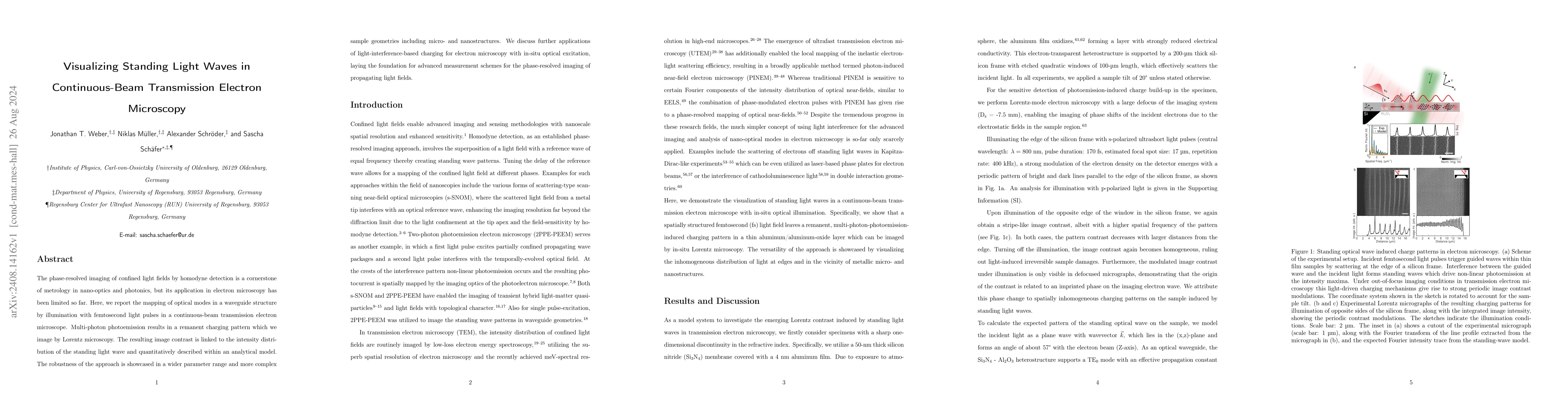
The phase-resolved imaging of confined light fields by homodyne detection is a cornerstone of metrology in nano-optics and photonics, but its application in electron microscopy has been limited so far. Here, we report the mapping of optical modes in a waveguide structure by illumination with femtosecond light pulses in a continuous-beam transmission electron microscope. Multi-photon photoemission results in a remanent charging pattern which we image by Lorentz microscopy. The resulting image contrast is linked to the intensity distribution of the standing light wave and quantitatively described within an analytical model. The robustness of the approach is showcased in a wider parameter range and more complex sample geometries including micro- and nanostructures. We discuss further applications of light-interference-based charging for electron microscopy with in-situ optical excitation, laying the foundation for advanced measurement schemes for the phase-resolved imaging of propagating light fields.
AI Key Findings
Get AI-generated insights about this paper's methodology, results, significance, and more — seven facets brought into focus.
Impact
Paper Details
Authors
PDF Preview
Citation Network
Current paper (gray), citations (green), references (blue)
Display is limited for performance on very large graphs.
Discussion 0