Publication
Metrics
AI Quick Summary
This paper demonstrates the use of table-top extreme ultraviolet (EUV) microscopy for label-free visualization of microorganisms, achieving high-resolution ptychographic imaging of fungi and bacteria. The study highlights the potential of the EUV spectral region for biological imaging, revealing intracellular features with sub-60 nm resolution.
Paper Preview
Abstract
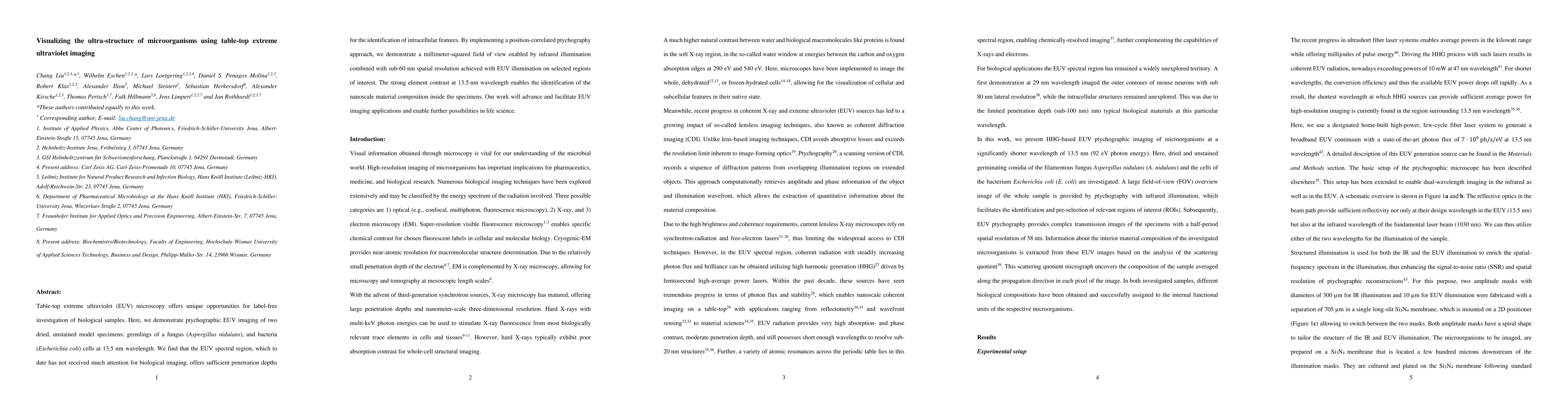
Table-top extreme ultraviolet (EUV) microscopy offers unique opportunities for label-free investigation of biological samples. Here, we demonstrate ptychographic EUV imaging of two dried, unstained model specimens: germlings of a fungus (Aspergillus nidulans), and bacteria (Escherichia coli) cells at 13.5 nm wavelength. We find that the EUV spectral region, which to date has not received much attention for biological imaging, offers sufficient penetration depths for the identification of intracellular features. By implementing a position-correlated ptychography approach, we demonstrate a millimeter-squared field of view enabled by infrared illumination combined with sub-60 nm spatial resolution achieved with EUV illumination on selected regions of interest. The strong element contrast at 13.5 nm wavelength enables the identification of the nanoscale material composition inside the specimens. Our work will advance and facilitate EUV imaging applications and enable further possibilities in life science.
AI Key Findings
Get AI-generated insights about this paper's methodology, results, significance, and more — seven facets brought into focus.
Impact
Paper Details
Authors
PDF Preview
Key Terms
Citation Network
Current paper (gray), citations (green), references (blue)
Display is limited for performance on very large graphs.
Discussion 0