X-ray diffraction with micrometer spatial resolution for highly absorbing samples
Publication
Metrics
AI Quick Summary
This paper presents a goniometer-based X-ray diffraction setup at P06 beamline of PETRA III that achieves micrometer spatial resolution for highly absorbing samples using photon energies above 35 keV. The method employs compound refractive lenses and enables 5D scans, demonstrating its application in determining local strain in martensitic steel and elemental distribution in high-Z materials within thin film solar cells.
Paper Preview
Abstract
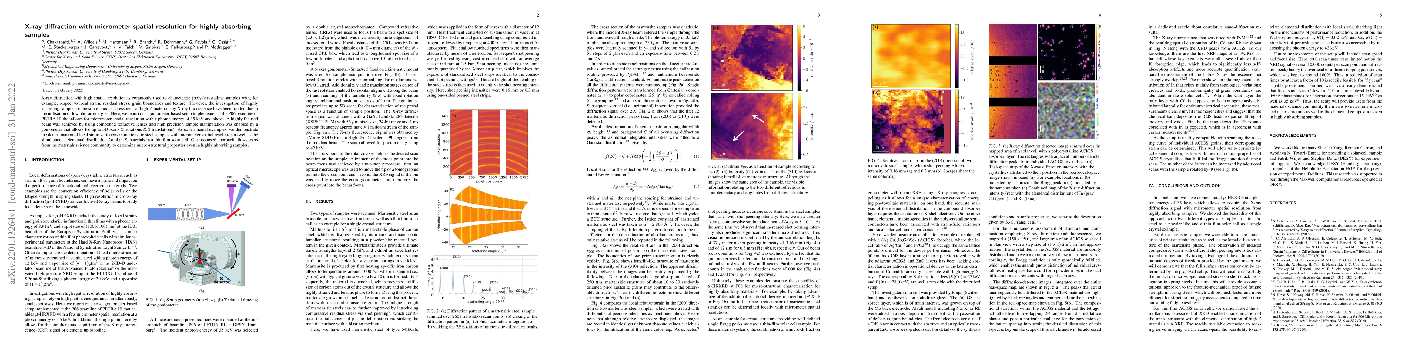
X-ray diffraction with high spatial resolution is commonly used to characterize (poly-)crystalline samples with, for example, respect to local strain, residual stress, grain boundaries and texture. However, the investigation of highly absorbing samples or the simultaneous assessment of high-Z materials by X-ray fluorescence have been limited due to the utilisation of low photon energies. Here, we report on a goniometer-based setup implemented at the P06 beamline of PETRA III that allows for micrometer spatial resolution with a photon energy of 35 keV and above. A highly focused beam was achieved by using compound refractive lenses and high precision sample manipulation was enabled by a goniometer that allows for up to 5D scans (3 rotations & 2 translations). As experimental examples, we demonstrate the determination of local strain variations in martensitic steel samples with micrometer spatial resolution as well as the simultaneous elemental distribution for high-Z materials in a thin film solar cell. Our proposed approach allows users from the materials science community to determine micro-structural properties even in highly absorbing samples.
AI Key Findings
Get AI-generated insights about this paper's methodology, results, significance, and more — seven facets brought into focus.
Impact
Paper Details
Authors
PDF Preview
Key Terms
Citation Network
Current paper (gray), citations (green), references (blue)
Display is limited for performance on very large graphs.
Discussion 0