Publication
Metrics
AI Quick Summary
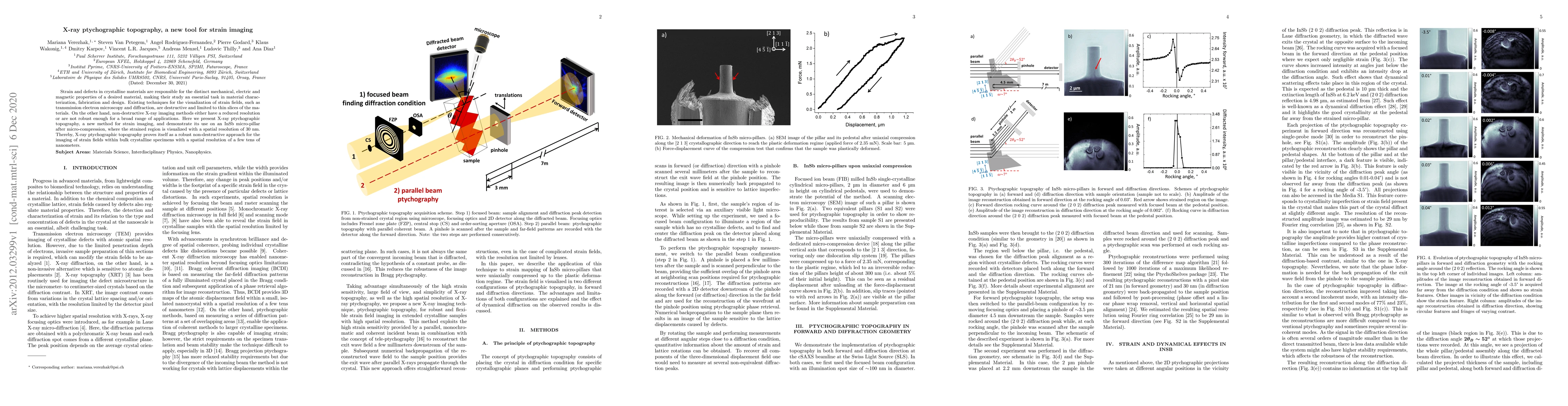
X-ray ptychographic topography is introduced as a non-destructive method for visualizing strain fields in crystalline materials with nanometer resolution. This technique demonstrates its effectiveness by imaging strain in an InSb micro-pillar, showing potential for broad applications in material characterization and design.
Paper Preview
Abstract
Strain and defects in crystalline materials are responsible for the distinct mechanical, electric and magnetic properties of a desired material, making their study an essential task in material characterization, fabrication and design. Existing techniques for the visualization of strain fields, such as transmission electron microscopy and diffraction, are destructive and limited to thin slices of the materials. On the other hand, non-destructive X-ray imaging methods either have a reduced resolution or are not robust enough for a broad range of applications. Here we present X-ray ptychographic topography, a new method for strain imaging, and demonstrate its use on an InSb micro-pillar after micro-compression, where the strained region is visualized with a spatial resolution of 30 nm. Thereby, X-ray ptychographic topography proves itself as a robust non-destructive approach for the imaging of strain fields within bulk crystalline specimens with a spatial resolution of a few tens of nanometers.
AI Key Findings
Get AI-generated insights about this paper's methodology, results, significance, and more — seven facets brought into focus.
Impact
Paper Details
Authors
PDF Preview
Key Terms
Citation Network
Current paper (gray), citations (green), references (blue)
Display is limited for performance on very large graphs.
Discussion 0