3D Blood Pulsation Maps

PDF

Publication

Published: Dec 11, 2025
Categories: cs.CV

Metrics

Source: ArXiv

Quick Actions

View PDF

Quick Answers

What methodology did the authors use?

The research employs a multi-view analysis of facial surfaces using FLAME-based modeling to extract physiological signals from skin color variations, combined with phase analysis from POS signals to infer blood perfusion patterns. More in Methodology →

What are the key results?

Significant variations in SNR were observed across different viewing angles and illumination conditions, with higher SNR near the camera center. — Phase inversion patterns were detected in facial regions connected to different parts of the arterial tree, validating physiological signal sources. More in Key Results →

Why is this work significant?

This research advances non-invasive physiological monitoring by enabling accurate ROI selection for blood perfusion analysis, which could improve diagnostic tools for cardiovascular health and real-time health monitoring systems. More in Significance →

What are the main limitations?

Motion artifacts significantly affect amplitude measurements, especially in dynamic facial regions. — The method's performance is compromised in areas with heavy facial hair or when subjects wear clothing. More in Limitations →

Paper Preview

Abstract

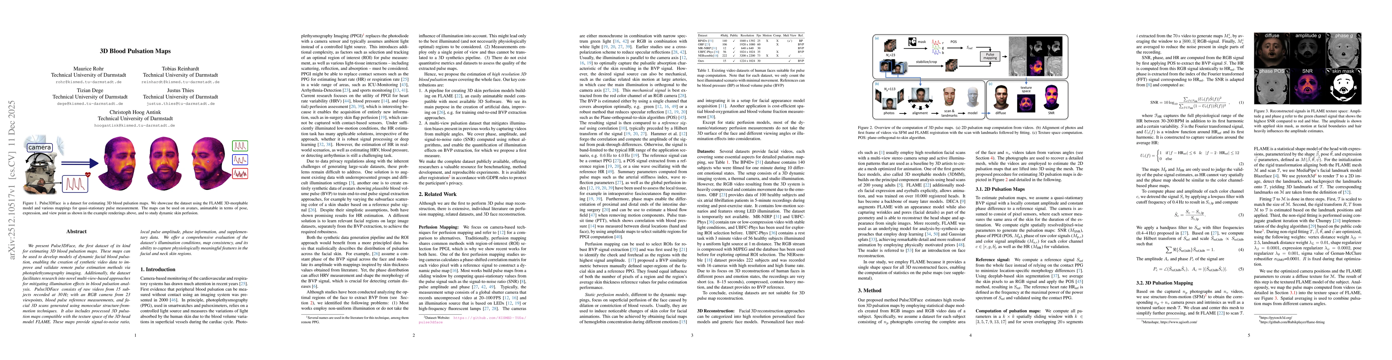
We present Pulse3DFace, the first dataset of its kind for estimating 3D blood pulsation maps. These maps can be used to develop models of dynamic facial blood pulsation, enabling the creation of synthetic video data to improve and validate remote pulse estimation methods via photoplethysmography imaging. Additionally, the dataset facilitates research into novel multi-view-based approaches for mitigating illumination effects in blood pulsation analysis. Pulse3DFace consists of raw videos from 15 subjects recorded at 30 Hz with an RGB camera from 23 viewpoints, blood pulse reference measurements, and facial 3D scans generated using monocular structure-from-motion techniques. It also includes processed 3D pulsation maps compatible with the texture space of the 3D head model FLAME. These maps provide signal-to-noise ratio, local pulse amplitude, phase information, and supplementary data. We offer a comprehensive evaluation of the dataset's illumination conditions, map consistency, and its ability to capture physiologically meaningful features in the facial and neck skin regions.

AI Key Findings

Generated Dec 13, 2025

Methodology — What approach did the authors take?

The research employs a multi-view analysis of facial surfaces using FLAME-based modeling to extract physiological signals from skin color variations, combined with phase analysis from POS signals to infer blood perfusion patterns.

Key Results — What are the main findings?

  • Significant variations in SNR were observed across different viewing angles and illumination conditions, with higher SNR near the camera center.
  • Phase inversion patterns were detected in facial regions connected to different parts of the arterial tree, validating physiological signal sources.
  • Amplitude maps showed increased values along facial borders and edges due to motion artifacts and cardiac effects.

Significance — Why does this research matter?

This research advances non-invasive physiological monitoring by enabling accurate ROI selection for blood perfusion analysis, which could improve diagnostic tools for cardiovascular health and real-time health monitoring systems.

Technical Contribution — What is the technical contribution?

The integration of FLAME-based surface modeling with phase analysis from POS signals provides a novel framework for extracting and analyzing physiological signals from facial skin color variations.

Novelty — What is new about this work?

This work uniquely combines multi-view facial analysis with phase information from POS signals, offering a more comprehensive understanding of blood perfusion patterns compared to traditional single-view methods.

Limitations — What are the limitations of this study?

  • Motion artifacts significantly affect amplitude measurements, especially in dynamic facial regions.
  • The method's performance is compromised in areas with heavy facial hair or when subjects wear clothing.

Future Work — What did the authors propose for future work?

  • Developing robust motion compensation algorithms to reduce artifacts in amplitude measurements.
  • Enhancing the model to handle occlusions caused by facial hair and clothing through improved segmentation techniques.

Paper Details

Paper ID: 2512.10517
Comments: 9 pages (without references), supplementals attached, waiting for publication. In order to access the dataset,see https://github.com/KISMED-TUDa/pulse3dface
Categories:
cs.CV

How to Cite This Paper

BibTeX
@article{antink2025d,
  title         = {3D Blood Pulsation Maps},
  author        = {Antink, Christoph Hoog and Thies, Justus and Rohr, Maurice and others},
  year          = {2025},
  eprint        = {2512.10517},
  archivePrefix = {arXiv},
  primaryClass  = {cs.CV},
}
APA 7
Antink, C., Thies, J., Rohr, M., Dege, T., & Reinhardt, T. (2025). 3D Blood Pulsation Maps. arXiv. https://arxiv.org/abs/2512.10517
MLA 9
Antink, Christoph Hoog, et al. "3D Blood Pulsation Maps." arXiv, 2025, arxiv.org/abs/2512.10517.

Comments (0)

This website uses cookies to ensure you get the best experience. Learn more