01
MethodologyHow they did it
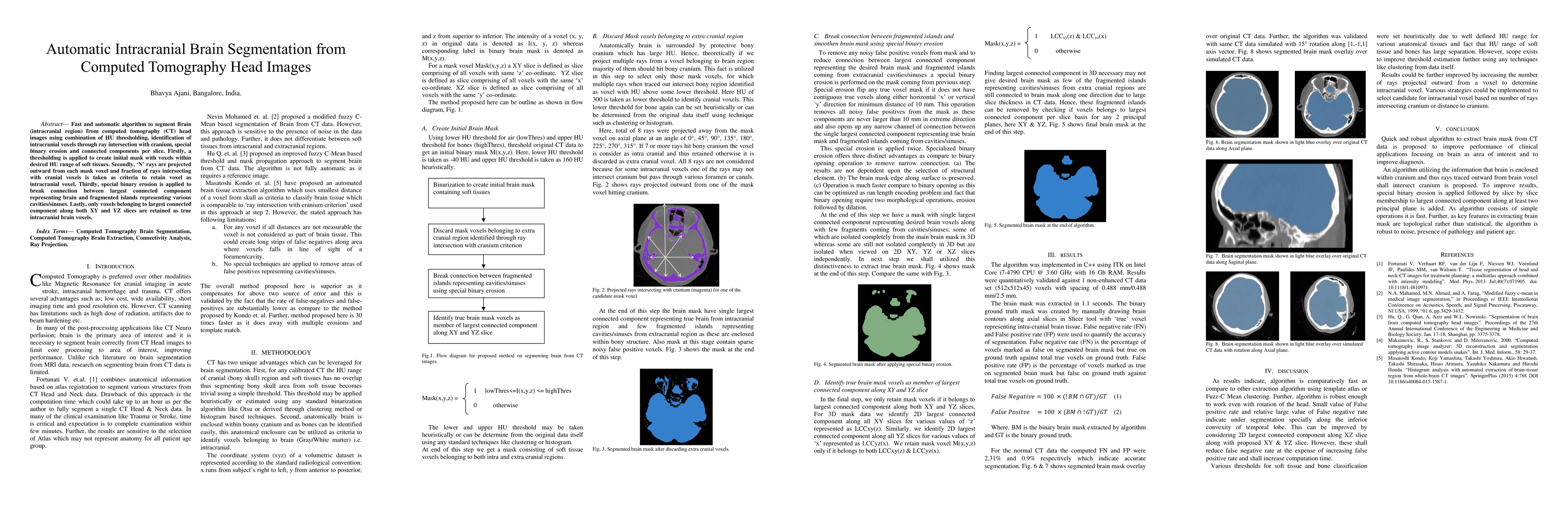
An algorithm utilizing the information that brain is enclosed within cranium and thus rays traced outward from brain voxel shall intersect cranium is proposed.
Discussion 0