Publication
Metrics
AI Quick Summary
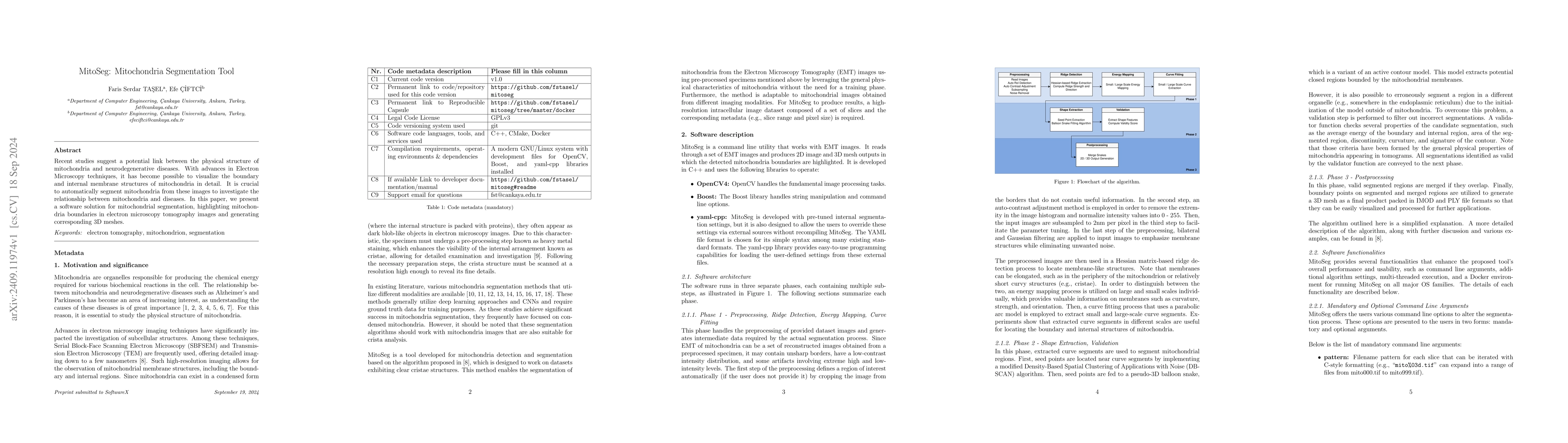
This paper introduces MitoSeg, a software tool designed to automatically segment mitochondria from electron microscopy tomography images, emphasizing mitochondria boundaries and generating detailed 3D meshes to explore the link between mitochondrial structure and neurodegenerative diseases.
Paper Preview
Abstract
Recent studies suggest a potential link between the physical structure of mitochondria and neurodegenerative diseases. With advances in Electron Microscopy techniques, it has become possible to visualize the boundary and internal membrane structures of mitochondria in detail. It is crucial to automatically segment mitochondria from these images to investigate the relationship between mitochondria and diseases. In this paper, we present a software solution for mitochondrial segmentation, highlighting mitochondria boundaries in electron microscopy tomography images and generating corresponding 3D meshes.
AI Key Findings
Get AI-generated insights about this paper's methodology, results, significance, and more — seven facets brought into focus.
Impact
Authors
PDF Preview
Citation Network
Current paper (gray), citations (green), references (blue)
Display is limited for performance on very large graphs.
Discussion 0