Publication
Metrics
AI Quick Summary
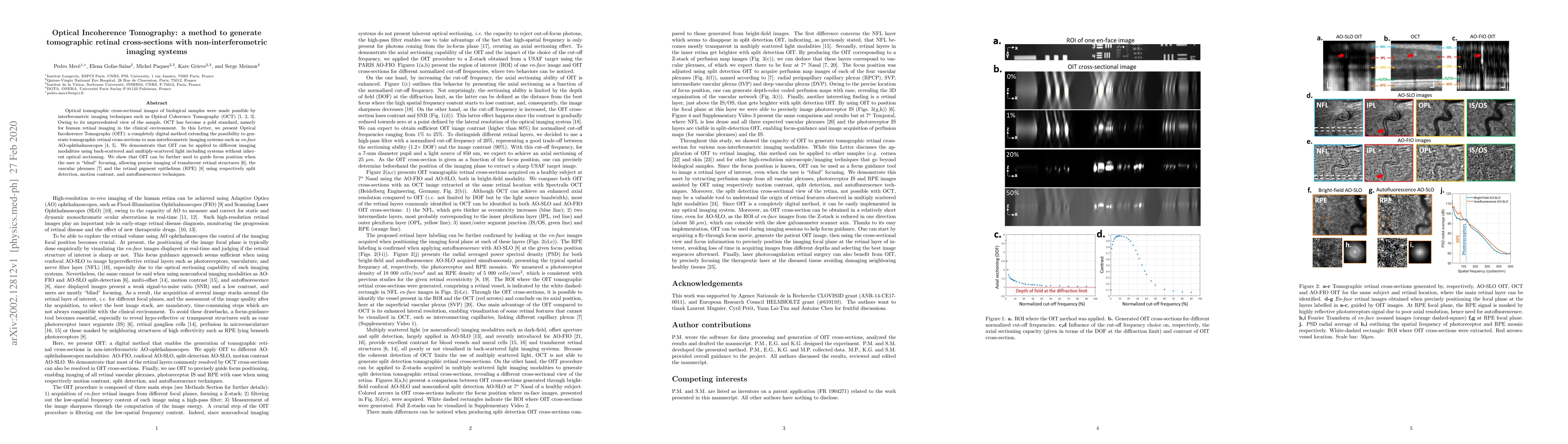
The research introduces Optical Incoherence Tomography (OIT), a method enabling tomographic retinal cross-sections using non-interferometric imaging systems like en-face AO-ophthalmoscopes. OIT can be applied to various imaging modalities, guiding focus for precise imaging of retinal structures, including vascular plexuses and retinal pigment epithelium.
Paper Preview
Abstract
Optical tomographic cross-sectional images of biological samples were made possible by interferometric imaging techniques such as Optical Coherence Tomography (OCT). Owing to its unprecedented view of the sample, OCT has become a gold standard, namely for human retinal imaging in the clinical environment. In this Letter, we present Optical Incoherence Tomography (OIT): a completely digital method extending the possibility to generate tomographic retinal cross-sections to non-interferometric imaging systems such as en-face AO-ophthalmoscopes. We demonstrate that OIT can be applied to different imaging modalities using back-scattered and multiply-scattered light including systems without inherent optical sectioning. We show that OIT can be further used to guide focus position when the user is "blind" focusing, allowing precise imaging of translucent retinal structures, the vascular plexuses and the retinal pigment epithelium using respectively split detection, motion contrast, and autofluorescence techniques.
AI Key Findings
Get AI-generated insights about this paper's methodology, results, significance, and more — seven facets brought into focus.
Impact
Paper Details
Authors
PDF Preview
Key Terms
Citation Network
Current paper (gray), citations (green), references (blue)
Display is limited for performance on very large graphs.
Discussion 0