ArxivLens

Publication

Published: Mar 04, 2026
Categories: cs.CV

Metrics

Source: ArXiv

Quick Answers

What methodology did the authors use?

Pre‑train a 3D masked autoencoder (MAE) on 5,000 prostate mpMRI patients (22,000 volumes, 1.8M slices) using T2w, DWI, and ADC modalities, then fine‑tune on 11 diverse downstream tasks (detection, grading, segmentation, localisation, volume estimation) across 3,000 patients, comparing against specialised and generalist foundation models. More in Methodology →

What are the key results?

ProFound‑Conv achieves the highest Dice (0.429±0.009) for prostate lesion segmentation and outperforms specialised UNet (0.416±0.004, p=0.001). — ProFound‑ViT attains the best anatomy segmentation Dice (0.931±0.001) and outperforms Rad‑Diag (0.921±0.001). More in Key Results →

Why is this work significant?

The study delivers a domain‑specialised vision foundation model that unifies multiple prostate imaging tasks, enabling expert‑level performance with fewer annotations and improving scalability for clinical deployment. More in Significance →

What are the main limitations?

Pre‑training and fine‑tuning demand extensive compute (10–14 days on high‑end GPUs). — Task performance varies across datasets due to domain shift and limited radiologist‑positive lesion data. More in Limitations →

Paper Preview

Abstract

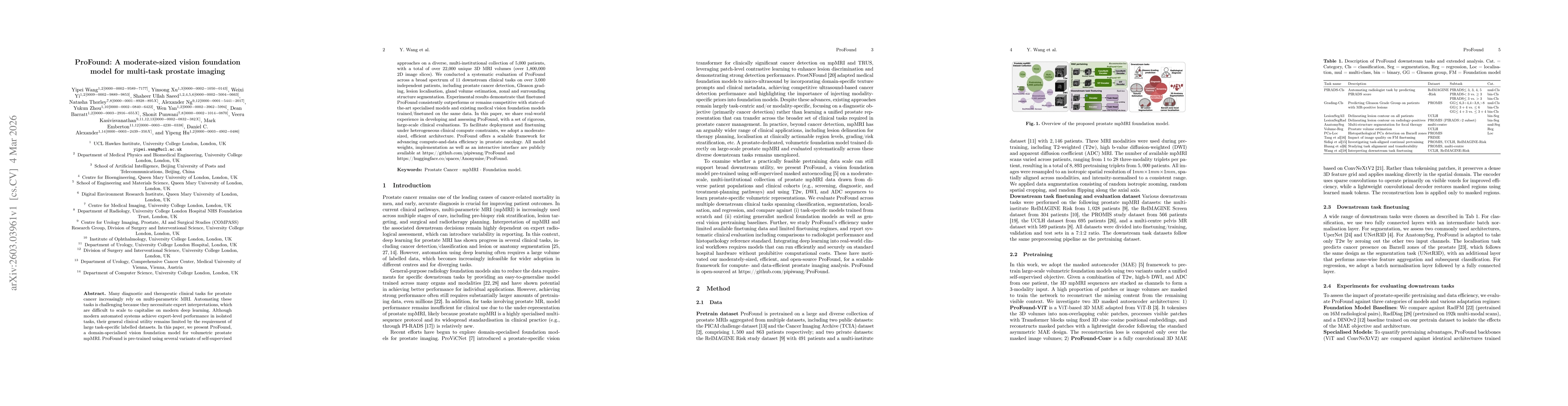
Many diagnostic and therapeutic clinical tasks for prostate cancer increasingly rely on multi-parametric MRI. Automating these tasks is challenging because they necessitate expert interpretations, which are difficult to scale to capitalise on modern deep learning. Although modern automated systems achieve expert-level performance in isolated tasks, their general clinical utility remains limited by the requirement of large task-specific labelled datasets. In this paper, we present ProFound, a domain-specialised vision foundation model for volumetric prostate mpMRI. ProFound is pre-trained using several variants of self-supervised approaches on a diverse, multi-institutional collection of 5,000 patients, with a total of over 22,000 unique 3D MRI volumes (over 1,800,000 2D image slices). We conducted a systematic evaluation of ProFound across a broad spectrum of $11$ downstream clinical tasks on over 3,000 independent patients, including prostate cancer detection, Gleason grading, lesion localisation, gland volume estimation, zonal and surrounding structure segmentation. Experimental results demonstrate that finetuned ProFound consistently outperforms or remains competitive with state-of-the-art specialised models and existing medical vision foundation models trained/finetuned on the same data.

Key Findings, in focus

Seven facets of this paper, analysed and brought into focus by AI.

AI-generated · verify against source Generated Mar 05, 2026
In focus · one-line synthesis

The study delivers a domain‑specialised vision foundation model that unifies multiple prostate imaging tasks, enabling expert‑level performance with fewer annotations and improving scalability for clinical deployment.

01
MethodologyHow they did it

Pre‑train a 3D masked autoencoder (MAE) on 5,000 prostate mpMRI patients (22,000 volumes, 1.8M slices) using T2w, DWI, and ADC modalities, then fine‑tune on 11 diverse downstream tasks (detection, grading, segmentation, localisation, volume estimation) across 3,000 patients, comparing against specialised and generalist foundation models.

02
ResultsWhat they found
  • ProFound‑Conv achieves the highest Dice (0.429±0.009) for prostate lesion segmentation and outperforms specialised UNet (0.416±0.004, p=0.001).
  • ProFound‑ViT attains the best anatomy segmentation Dice (0.931±0.001) and outperforms Rad‑Diag (0.921±0.001).
  • In low‑label settings ProFound reaches Dice 0.276 with only 32 fine‑tuning cases, consistently surpassing UNet across all low‑data regimes.
03
SignificanceWhy it matters

The study delivers a domain‑specialised vision foundation model that unifies multiple prostate imaging tasks, enabling expert‑level performance with fewer annotations and improving scalability for clinical deployment.

04
Technical ContributionsWhat's new technically

A 3D masked autoencoder foundation model (ViT and ConvNeXt variants) trained on multi‑modality prostate mpMRI, demonstrating superior representation learning for multi‑task prostate imaging.

05
NoveltyWhat sets it apart

First moderate‑sized, prostate‑specific vision foundation model that combines self‑supervised MAE pretraining on diverse multi‑parametric MRI and extensive downstream evaluation, outperforming generalist models.

06
LimitationsWhat to be cautious about
  • Pre‑training and fine‑tuning demand extensive compute (10–14 days on high‑end GPUs).
  • Task performance varies across datasets due to domain shift and limited radiologist‑positive lesion data.
07
Future WorkWhere it goes next
  • Expand the pre‑training corpus with larger, higher‑quality multi‑institutional mpMRI datasets.
  • Investigate continual pre‑training and task‑aligned adaptation to further boost downstream accuracy and robustness.

Paper Details

Paper ID: 2603.03961
Categories:
cs.CV

How to Cite This Paper

Reference manager: RIS EndNote


Discussion 0

This website uses cookies to ensure you get the best experience. Learn more