01
MethodologyHow they did it
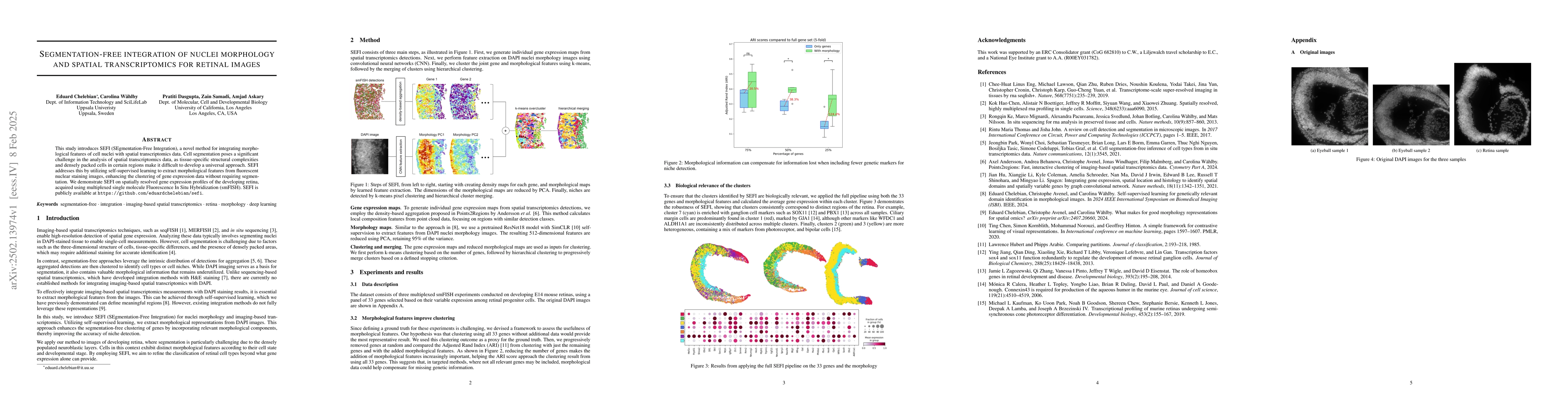
SEFI (Segmentation-Free Integration) method extracts morphological features from fluorescent nuclear staining images using self-supervised learning, enhancing clustering of gene expression data without cell segmentation. It consists of three main steps: generating individual gene expression maps, feature extraction on DAPI nuclei morphology images using CNN, and clustering the joint gene and morphological features using k-means followed by hierarchical clustering.
Discussion 0