Academic Profile
Statistics
Similar Authors
Papers on arXiv
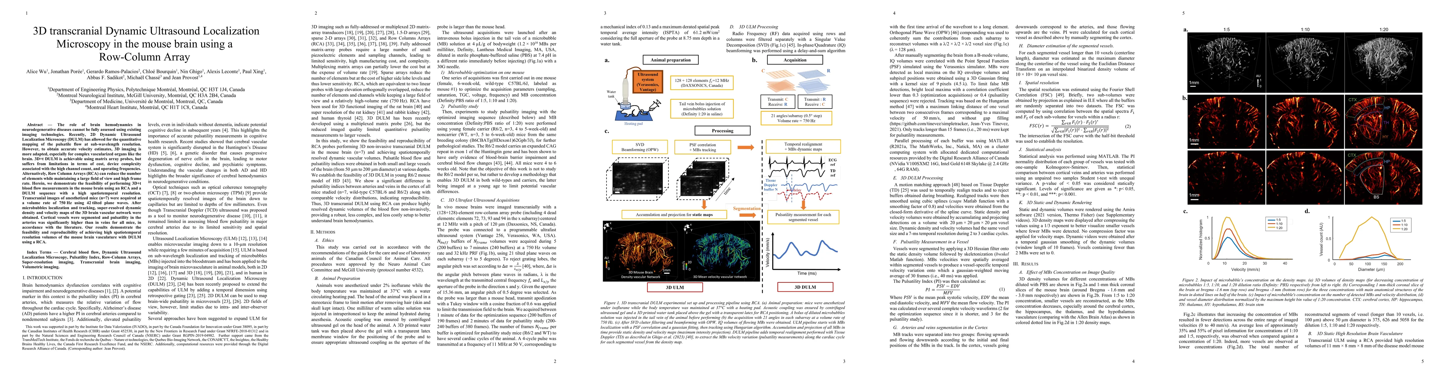
The role of brain hemodynamics in neurodegenerative diseases cannot be fully assessed using existing imaging technologies. Recently, 2D Dynamic Ultrasound Localization Microscopy (DULM) has allowed ...
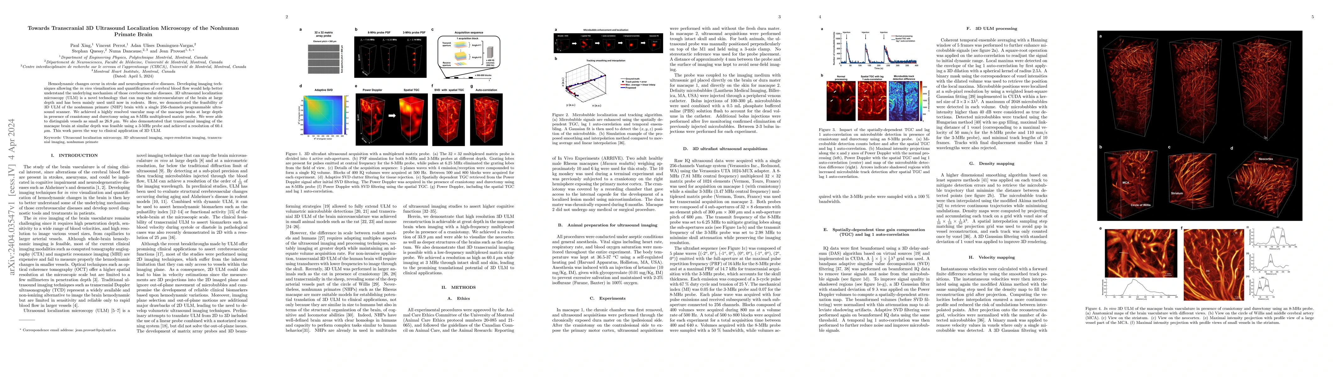
Hemodynamic changes occur in stroke and neurodegenerative diseases. Developing imaging techniques allowing the in vivo visualization and quantification of cerebral blood flow would help better under...
Color Doppler echocardiography enables visualization of blood flow within the heart. However, the limited frame rate impedes the quantitative assessment of blood velocity throughout the cardiac cycl...
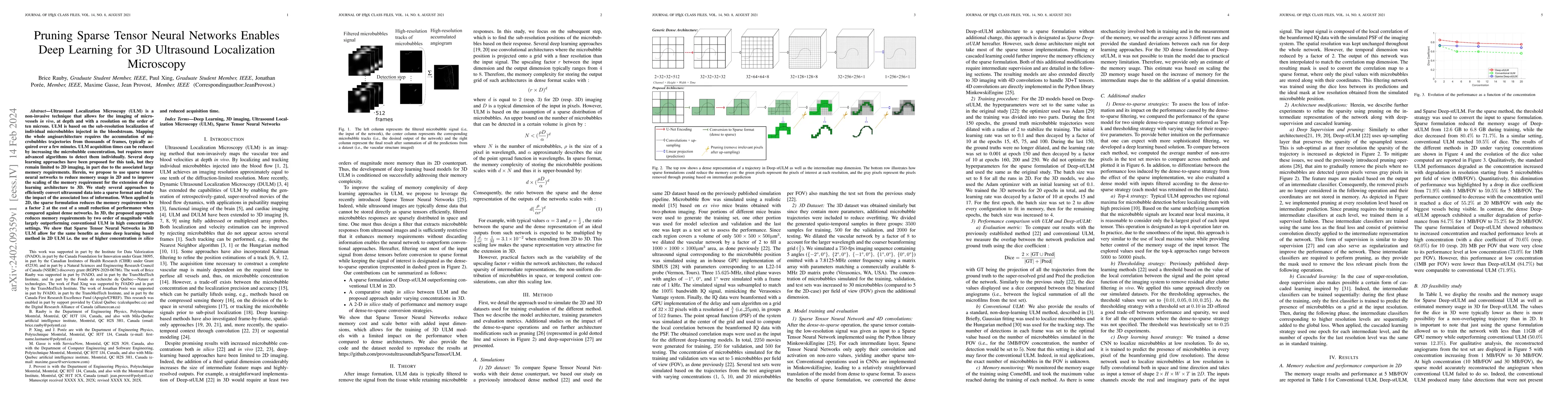
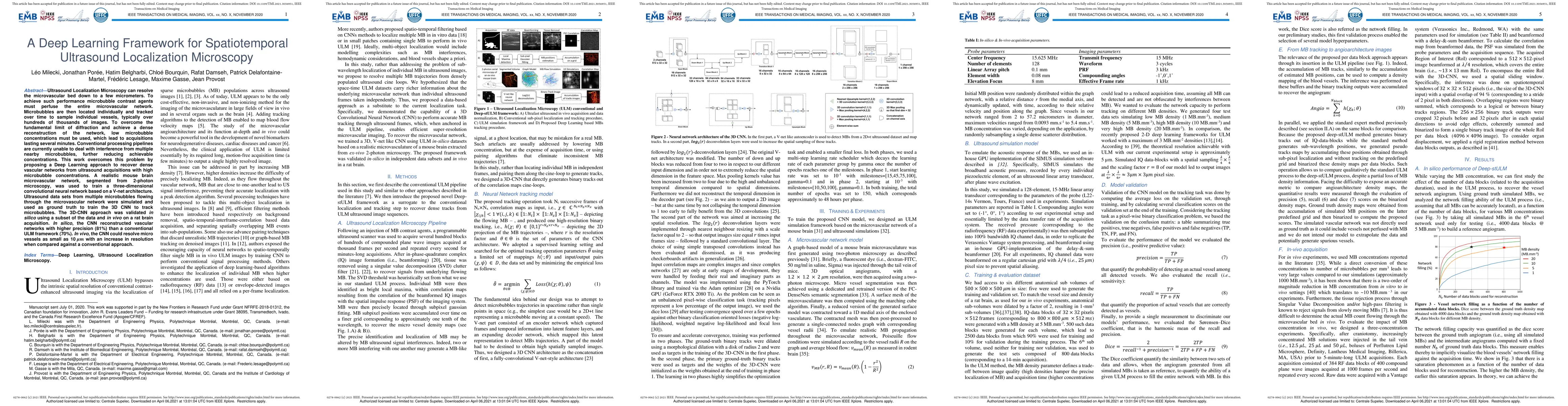
Ultrasound Localization Microscopy (ULM) is a non-invasive technique that allows for the imaging of micro-vessels in vivo, at depth and with a resolution on the order of ten microns. ULM is based on...
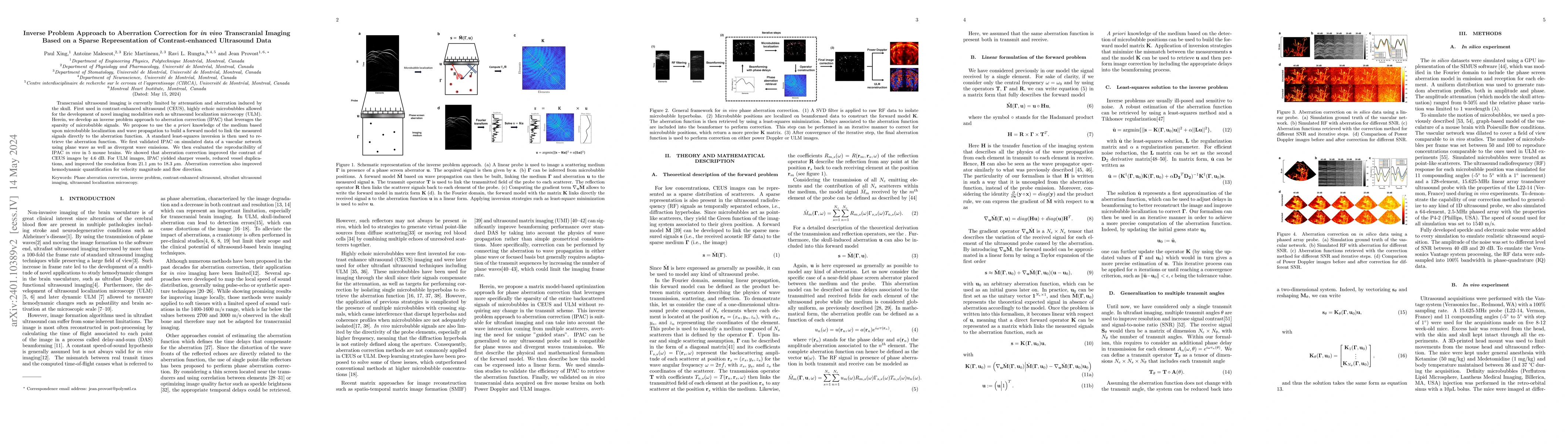
Transcranial ultrasound imaging is currently limited by attenuation and aberration induced by the skull. First used in contrast-enhanced ultrasound (CEUS), highly echoic microbubbles allowed for the...
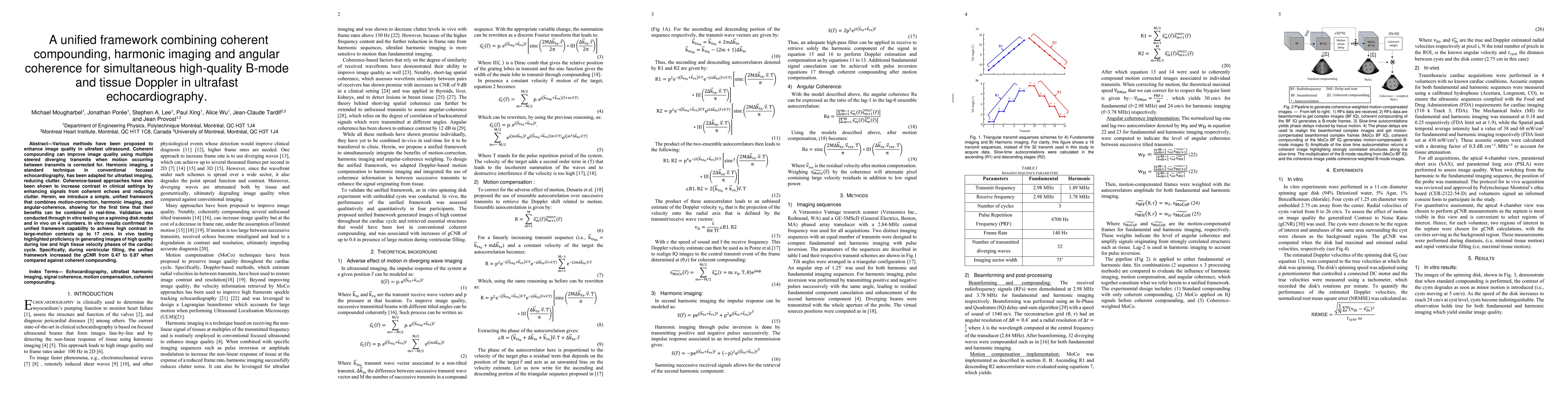
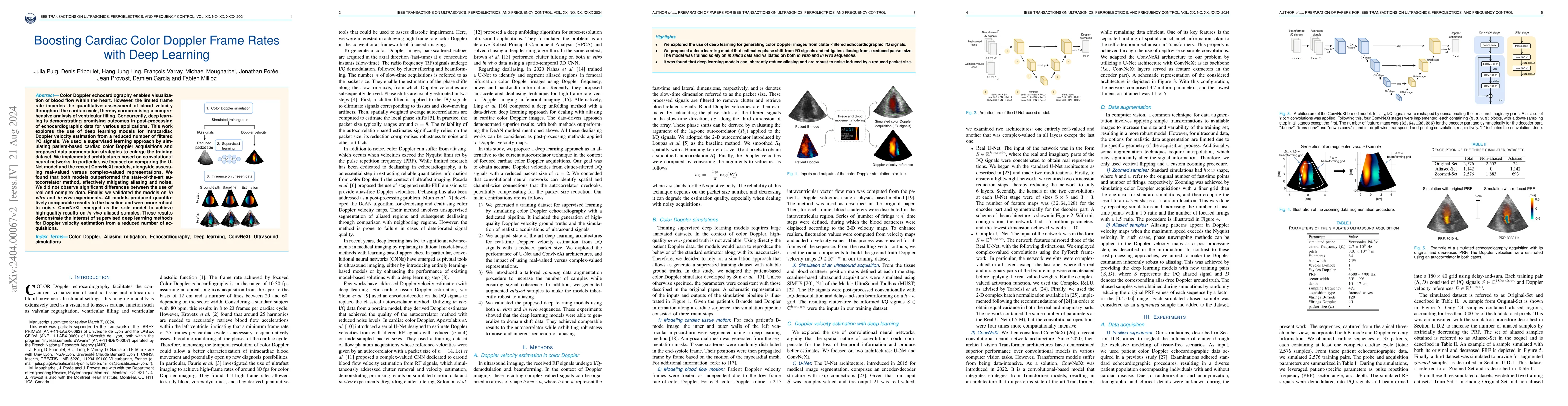
Various methods have been proposed to enhance image quality in ultrafast ultrasound. Coherent compounding can improve image quality using multiple steered diverging transmits when motion occurring b...
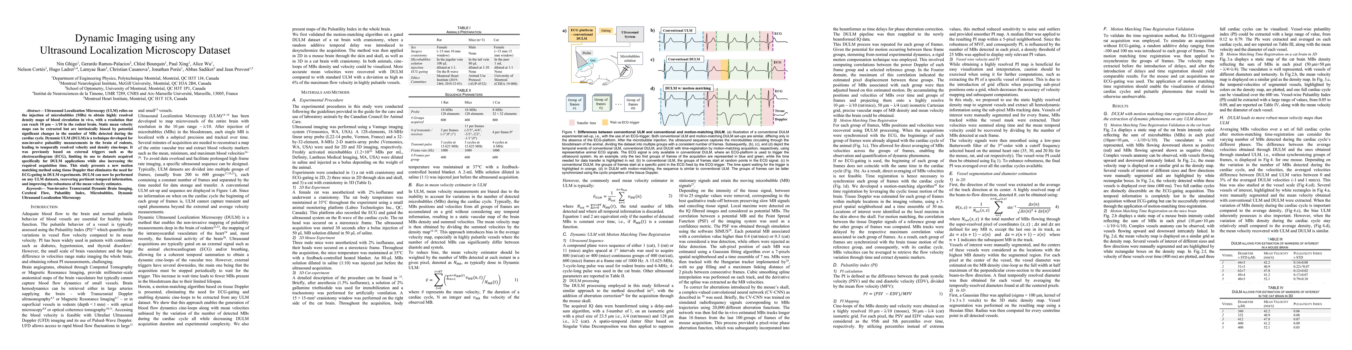
Ultrasound Localization Microscopy (ULM) relies on the injection of microbubbles (MBs) to obtain highly resolved density maps of blood circulation in vivo, with a resolution that can reach 10 {\mu}m...
Ultrasound Localization Microscopy (ULM) has recently enabled the mapping of the cerebral vasculature in vivo with a resolution ten times smaller than the wavelength used, down to ten microns. Howev...
The development of neurologically active drugs faces a challenge in treating brain diseases due to the blood-brain barrier's impermeability. High-intensity focused ultrasound can open the barrier in...
Ultrasound Localization Microscopy can resolve the microvascular bed down to a few micrometers. To achieve such performance microbubble contrast agents must perfuse the entire microvascular network....
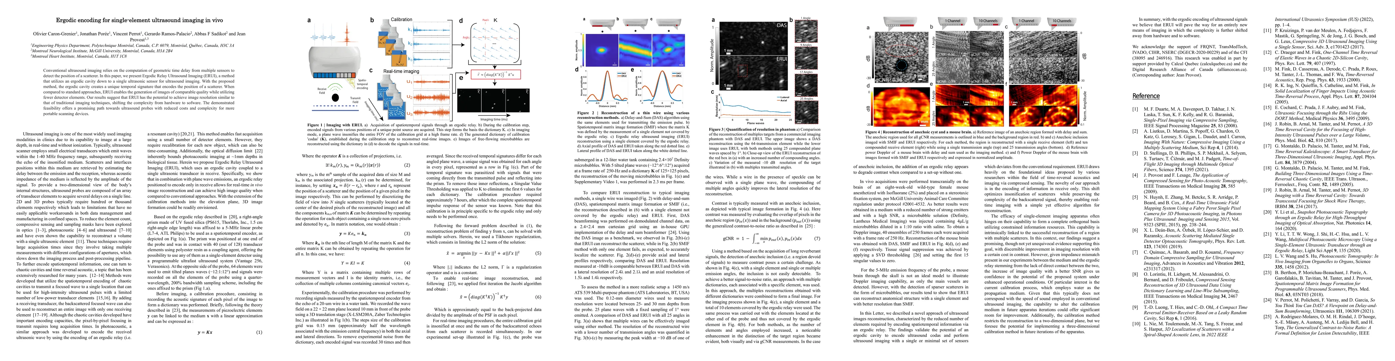
Conventional ultrasound imaging relies on the computation of geometric time delay from multiple sensors to detect the position of a scatterer. In this paper, we present Ergodic Relay Ultrasound Imag...
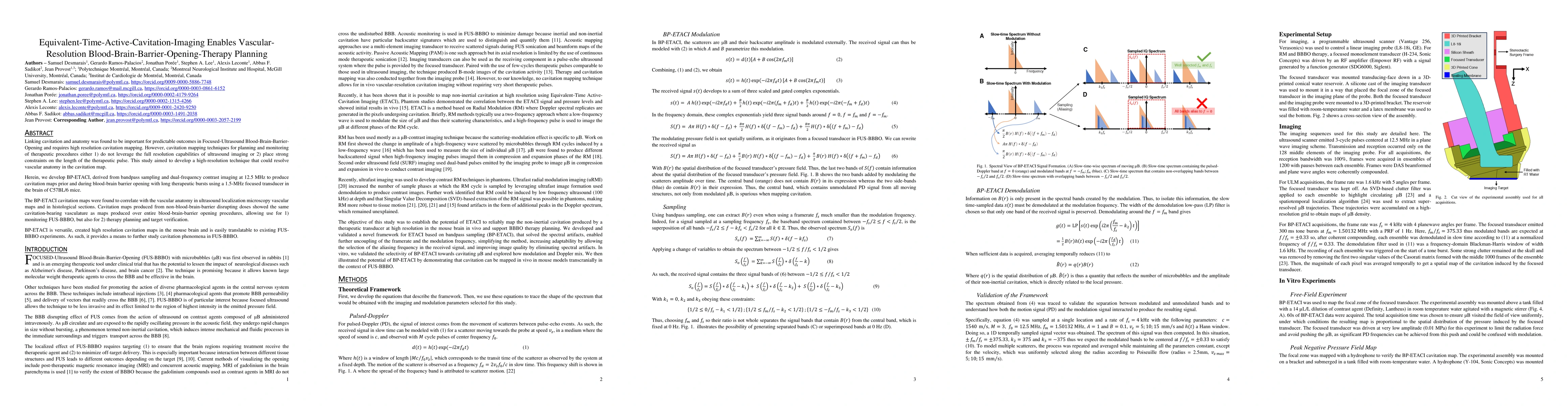
Linking cavitation and anatomy was found to be important for predictable outcomes in Focused-Ultrasound Blood-Brain-Barrier-Opening and requires high resolution cavitation mapping. However, cavitati...
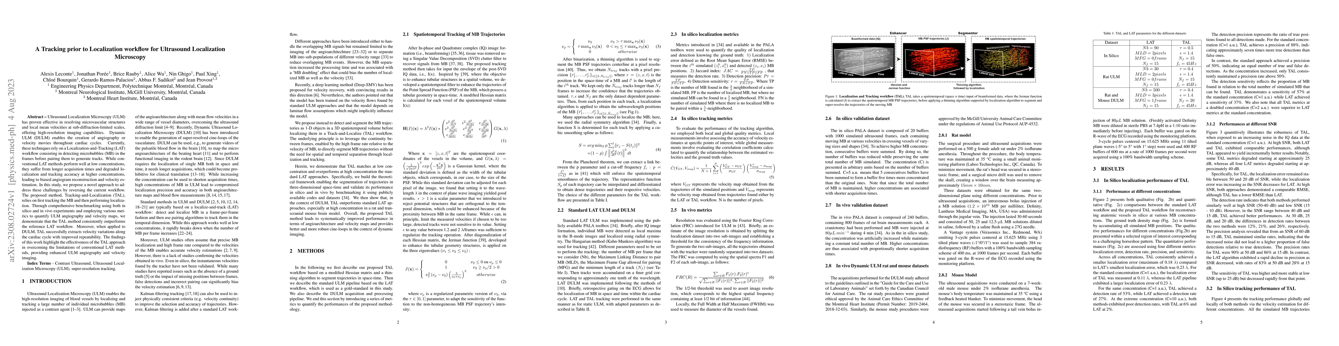
Ultrasound Localization Microscopy (ULM) has proven effective in resolving microvascular structures and local mean velocities at sub-diffraction-limited scales, offering high-resolution imaging capa...
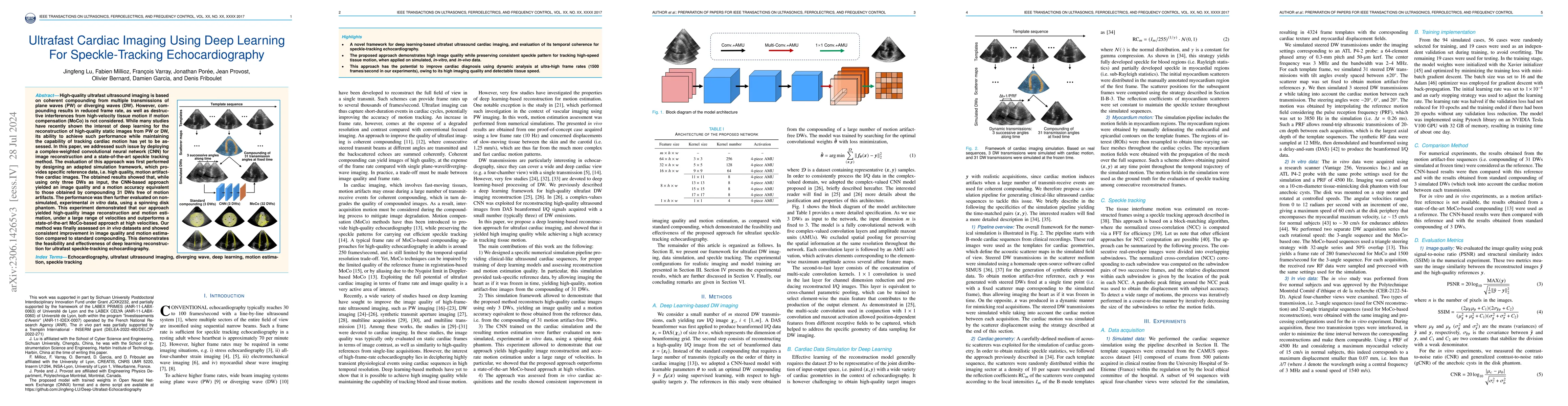
High-quality ultrafast ultrasound imaging is based on coherent compounding from multiple transmissions of plane waves (PW) or diverging waves (DW). However, compounding results in reduced frame rate...
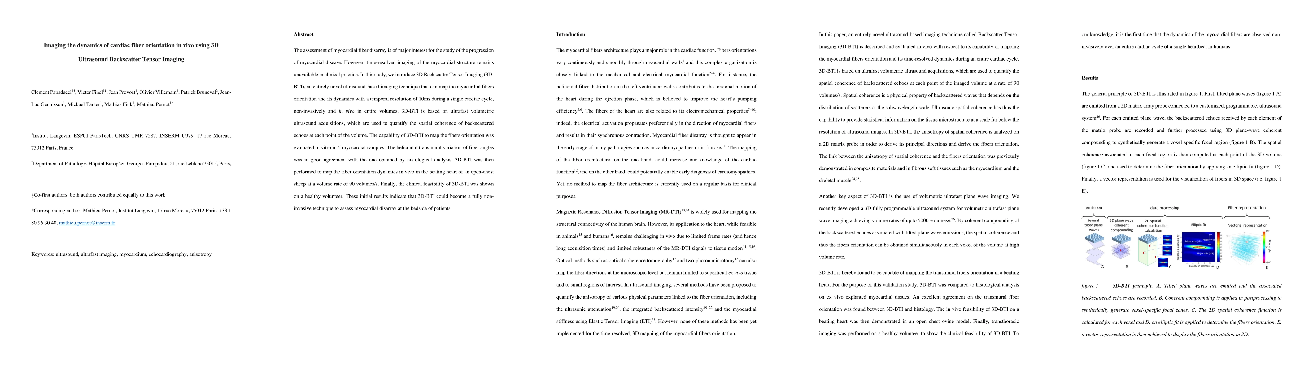
The assessment of myocardial fiber disarray is of major interest for the study of the progression of myocardial disease. However, time-resolved imaging of the myocardial structure remains unavailabl...
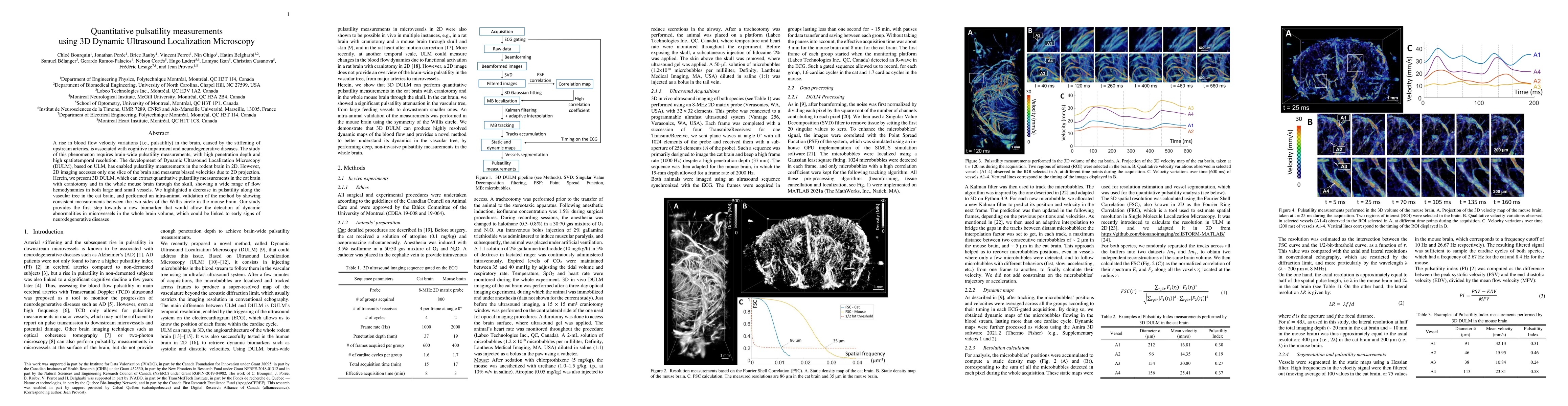
A rise in blood flow velocity variations (i.e., pulsatility) in the brain, caused by the stiffening of upstream arteries, is associated with cognitive impairment and neurodegenerative diseases. The ...
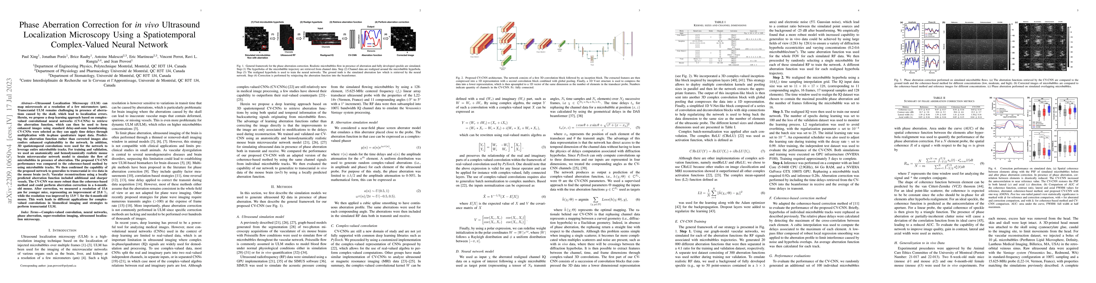
Ultrasound Localization Microscopy (ULM) can map microvessels at a resolution of a few micrometers (\mu m). Transcranial ULM remains challenging in presence of aberrations caused by the skull, which...
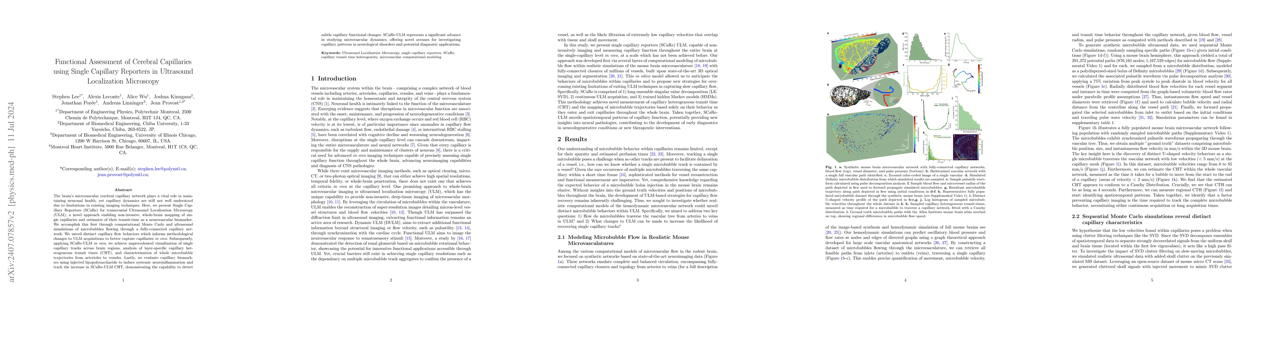
The brain's microvascular cerebral capillary network plays a vital role in maintaining neuronal health, yet capillary dynamics are still not well understood due to limitations in existing imaging tech...
The-Bodega is a Matlab-based toolbox for simulating ground-truth datasets for Ultrasound Localization Microscopy (ULM)-a super resolution imaging technique that resolves microvessels by systematically...
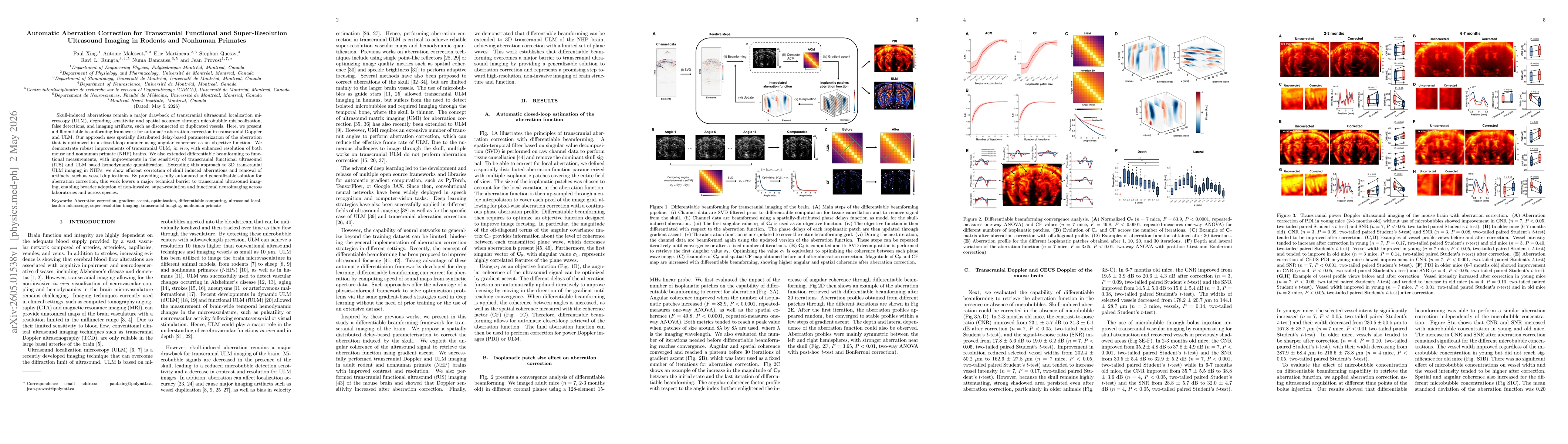
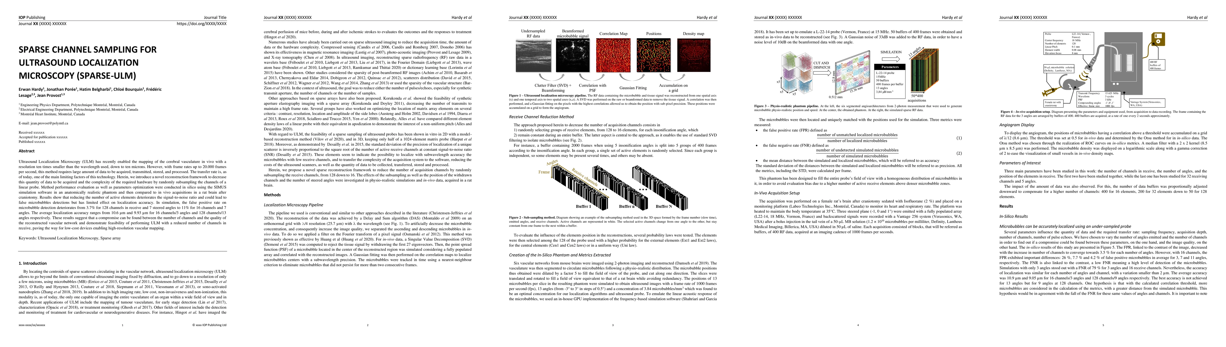
Skull-induced aberrations remain a major drawback of transcranial ultrasound localization microscopy (ULM), degrading sensitivity and spatial accuracy through microbubble mislocalization, false detect...
Ultrasound Localization Microscopy (ULM) enables microscopic imaging of the cerebral microvasculature in vivo, but relies on a multi-stage processing pipeline in which acquisition settings and reconst...