ArxivLens

Quick Summary:

This paper introduces a new motion matching method using tissue Doppler for Dynamic Ultrasound Localization Microscopy (DULM), eliminating the need for ECG-gating. This advancement allows DULM to be applied to any Ultrasound Localization Microscopy dataset, enhancing temporal resolution and robustness of velocity measurements.

arXiv Id: 2311.00648
Comments: 11 pages, 6 figures, 4 tables
Date Published: 2023-11-02
Date Updated: 2023-11-02

Categories:

Key Terms (TF-IDF):

brain (0.256)
resolved (0.222)
velocity (0.216)
maps (0.197)
ultrasound (0.182)
mean (0.170)
datasets (0.166)
doppler (0.165)
injection (0.157)
intrinsically (0.156)
loops (0.152)
acquired (0.146)
acquisition (0.145)
biased (0.144)
density (0.141)
microscopy (0.140)
limiting (0.137)
cycle (0.134)
localization (0.132)
reach (0.131)
License: http://creativecommons.org/licenses/by/4.0/

Summary:

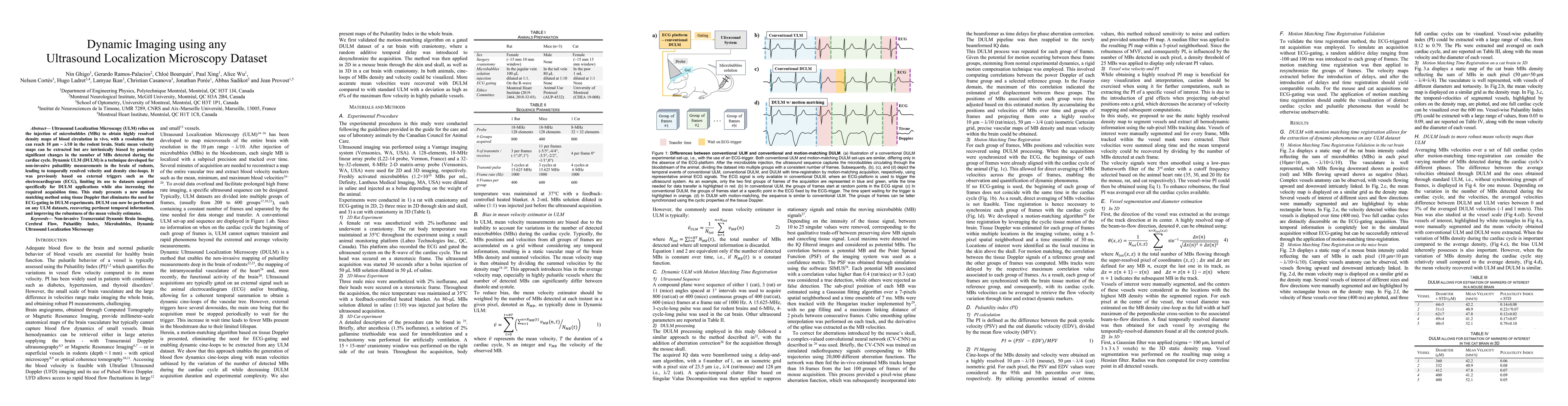
Ultrasound Localization Microscopy (ULM) relies on the injection of microbubbles (MBs) to obtain highly resolved density maps of blood circulation in vivo, with a resolution that can reach 10 {\mu}m ~ {\lambda}/10 in the rodent brain. Static mean velocity maps can be extracted but are intrinsically biased by potential significant changes in the number of MBs detected during the cardiac cycle. Dynamic ULM (DULM) is a technique developed for non-invasive pulsatility measurements in the brain of rodents, leading to temporally resolved velocity and density cine-loops. It was previously based on external triggers such as the electrocardiogram (ECG), limiting its use to datasets acquired specifically for DULM applications while also increasing the required acquisition time. This study presents a new motion matching method using tissue Doppler that eliminates the need for ECG-gating in DULM experiments. DULM can now be performed on any ULM datasets, recovering pertinent temporal information, and improving the robustness of the mean velocity estimates.

Citation & Reference Network Visualization

This graph shows how this paper is connected to other research. The current paper is shown in dark gray, papers that cite it are in green, and papers it references are in blue. You can:

Current Paper
Papers citing this work (Citations)
Papers cited by this work (References)

• Hover over any node to see the paper title

• Click a node to view that paper's details

• Drag nodes to rearrange the graph

• Use the zoom controls (bottom right) to zoom in/out

Citations and References

Title Authors Year Actions
3D transcranial Dynamic Ultrasound Localization Microscopy in the mouse brain using a Row-Column Array
Alice Wu, Jonathan Por'ee, Gerardo Ramos-Palacios, Chloé Bourquin, Nin Ghigo, Alexis Leconte, Paul Xing, Abbas Sadikot, Michael Chass'e, Jean Provost
2024
View Paper
Inverse Problem Approach to Aberration Correction for in vivo Transcranial Imaging Based on a Sparse Representation of Contrast-enhanced Ultrasound Data.
Paul Xing, Antoine Malescot, Éric Martineau, Ravi L. Rungta, Jean Provost
2024
View Paper

Similar Papers

Ultrasound matrix imaging for transcranial in-vivo localization microscopy

Mathias Fink, Olivier Couture, Flavien Bureau, Alexandre Aubry, Louise Denis, Antoine Coudert

Published: 2024-10-18

Quantitative pulsatility measurements using 3D Dynamic Ultrasound Localization Microscopy

Jonathan Porée, Gerardo Ramos-Palacios, Chloé Bourquin, Nin Ghigo, Jean Provost, Vincent Perrot, Brice Rauby, Christian Casanova, Nelson Cortés, Hugo Ladret, Lamyae Ikan, Hatim Belgharbi, Frédéric Lesage, Samuel Bélanger

Published: 2023-03-28

Geometric Ultrasound Localization Microscopy

Raphael Sznitman, Christopher Hahne

Published: 2023-07-19

Comments (0)

This website uses cookies to ensure you get the best experience. Learn more