Quantitative pulsatility measurements using 3D Dynamic Ultrasound Localization Microscopy
Publication
Metrics
AI Quick Summary
This paper introduces 3D Dynamic Ultrasound Localization Microscopy (DULM) for quantitative pulsatility measurements in the brain, addressing limitations of 2D DULM. The method enables comprehensive hemodynamic analysis in both cat and mouse brains, revealing decreased pulsatility along the vascular tree and validating measurements across different brain regions.
Paper Preview
Abstract
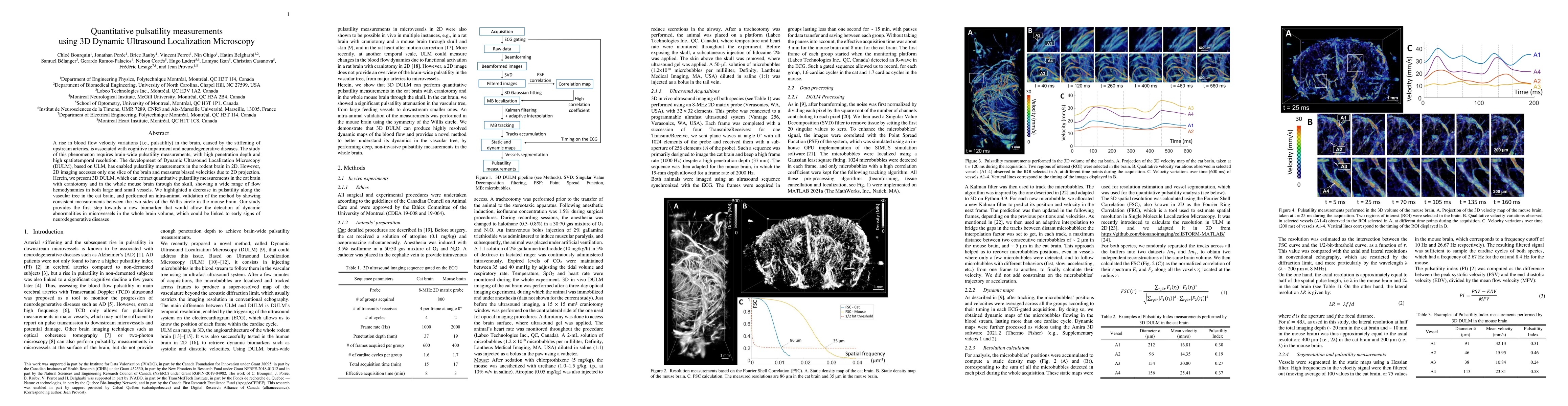
A rise in blood flow velocity variations (i.e., pulsatility) in the brain, caused by the stiffening of upstream arteries, is associated with cognitive impairment and neurodegenerative diseases. The study of this phenomenon requires brain-wide pulsatility measurements, with high penetration depth and high spatiotemporal resolution. The development of Dynamic Ultrasound Localization Microscopy (DULM), based on ULM, has enabled pulsatility measurements in the rodent brain in 2D. However, 2D imaging accesses only one slice of the brain and measures biased velocities due to 2D projection. Herein, we present 3D DULM, which can extract quantitative pulsatility measurements in the cat brain with craniotomy and in the whole mouse brain through the skull, showing a wide range of flow hemodynamics in both large and small vessels. We highlighted a decrease in pulsatility along the vascular tree in the cat brain, and performed an intra-animal validation of the method by showing consistent measurements between the two sides of the Willis circle in the mouse brain. Our study provides the first step towards a new biomarker that would allow the detection of dynamic abnormalities in microvessels in the whole brain volume, which could be linked to early signs of neurodegenerative diseases.
AI Key Findings
Get AI-generated insights about this paper's methodology, results, significance, and more — seven facets brought into focus.
Impact
Paper Details
Authors
PDF Preview
Key Terms
Citation Network
Current paper (gray), citations (green), references (blue)
Display is limited for performance on very large graphs.
Discussion 0