Academic Profile
Statistics
Similar Authors
Papers on arXiv
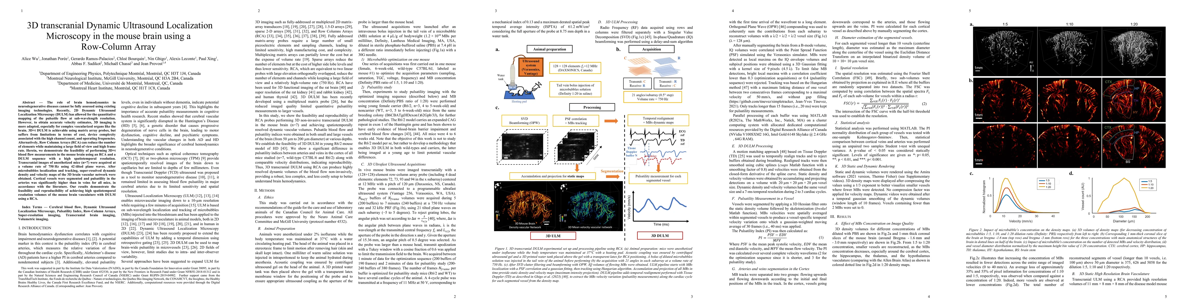
The role of brain hemodynamics in neurodegenerative diseases cannot be fully assessed using existing imaging technologies. Recently, 2D Dynamic Ultrasound Localization Microscopy (DULM) has allowed ...
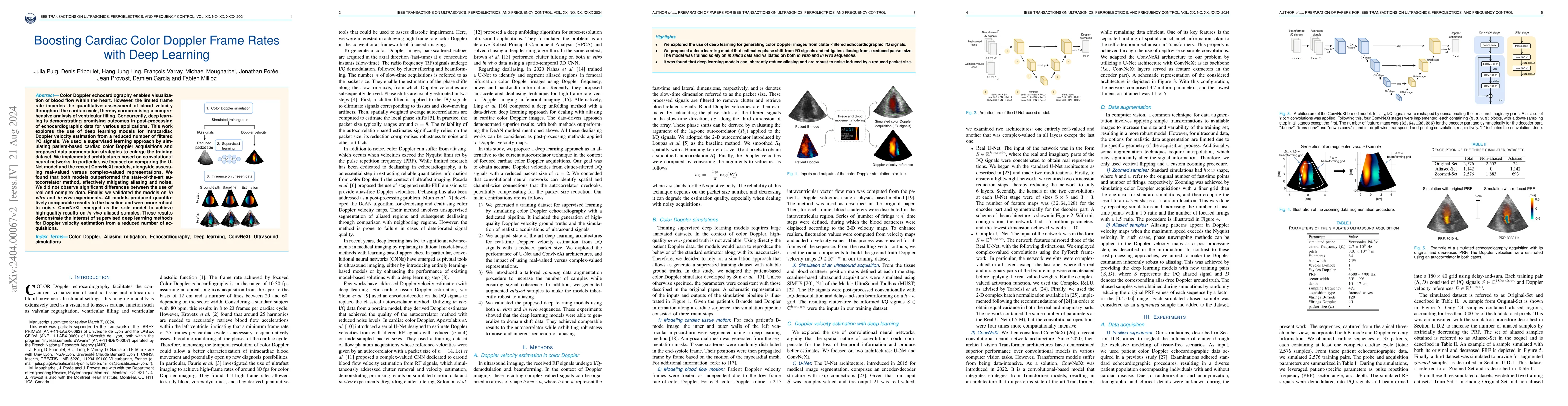
Color Doppler echocardiography enables visualization of blood flow within the heart. However, the limited frame rate impedes the quantitative assessment of blood velocity throughout the cardiac cycl...
Ultrasound Localization Microscopy (ULM) is a non-invasive technique that allows for the imaging of micro-vessels in vivo, at depth and with a resolution on the order of ten microns. ULM is based on...
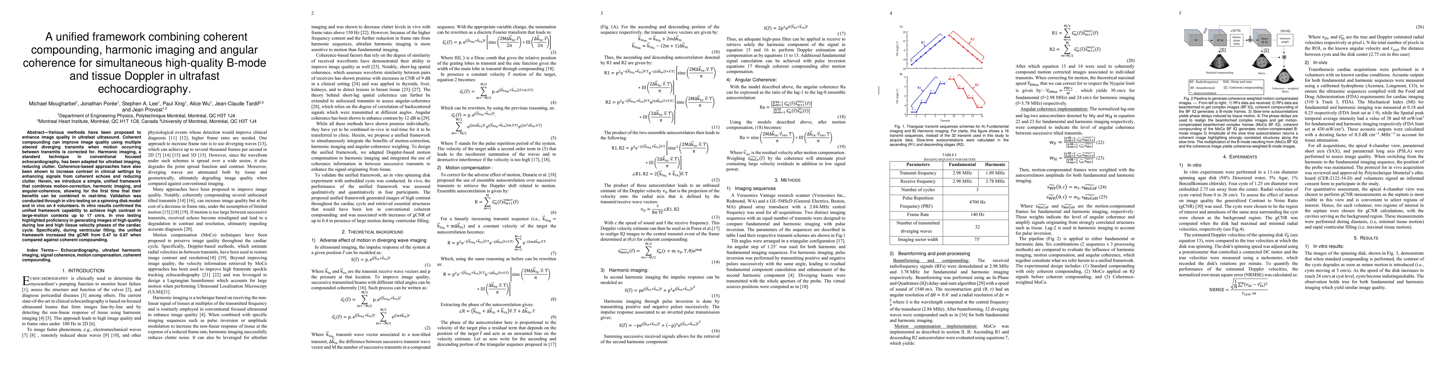
Various methods have been proposed to enhance image quality in ultrafast ultrasound. Coherent compounding can improve image quality using multiple steered diverging transmits when motion occurring b...
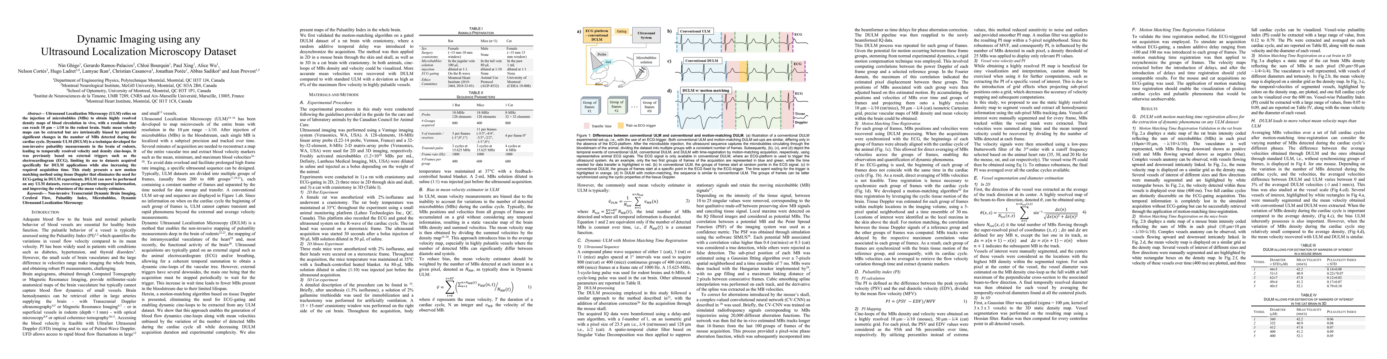
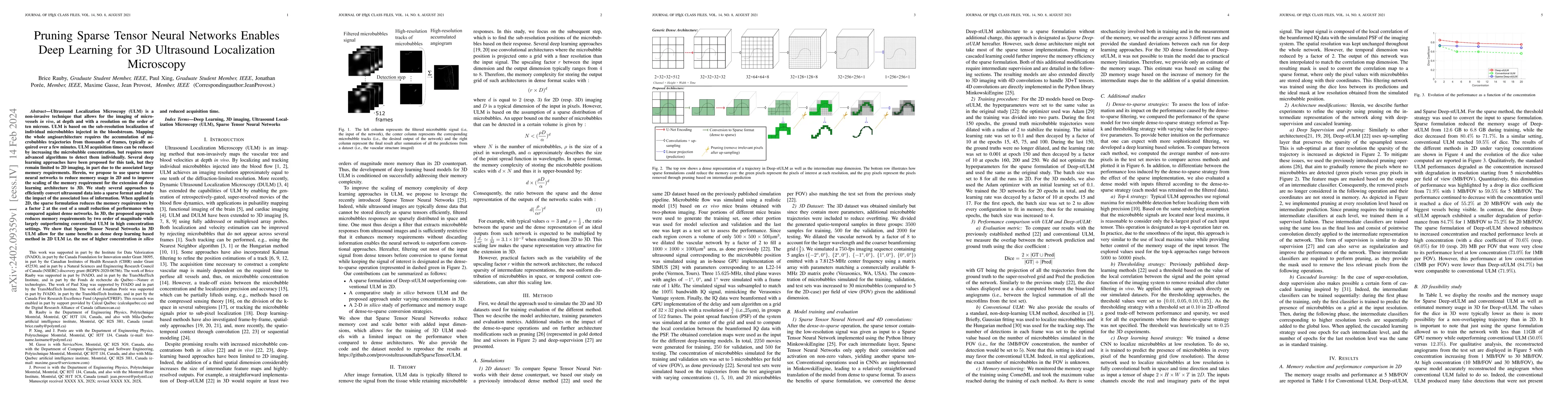
Ultrasound Localization Microscopy (ULM) relies on the injection of microbubbles (MBs) to obtain highly resolved density maps of blood circulation in vivo, with a resolution that can reach 10 {\mu}m...
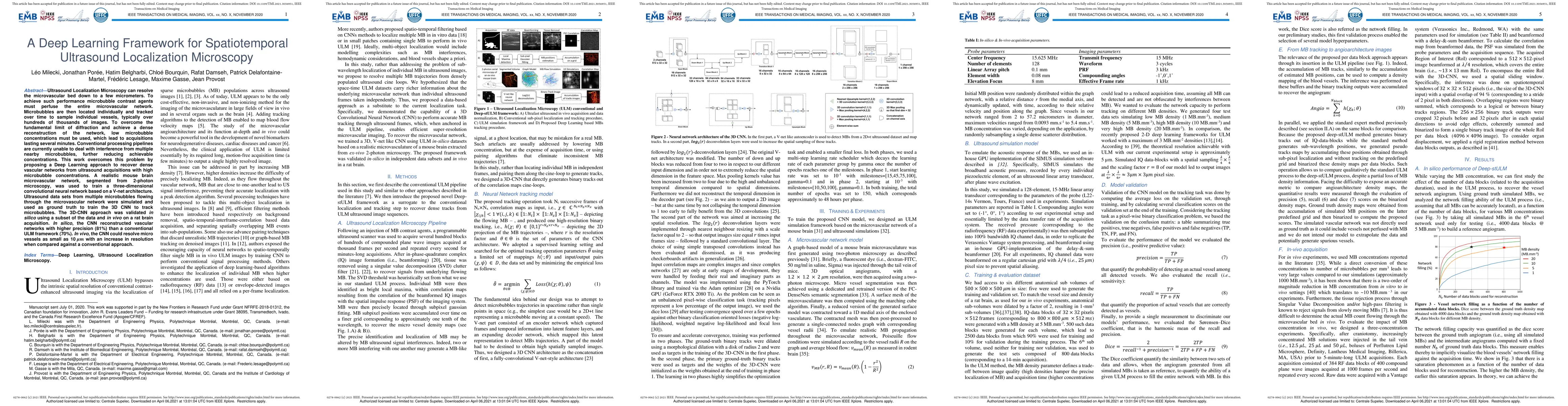
Ultrasound Localization Microscopy can resolve the microvascular bed down to a few micrometers. To achieve such performance microbubble contrast agents must perfuse the entire microvascular network....
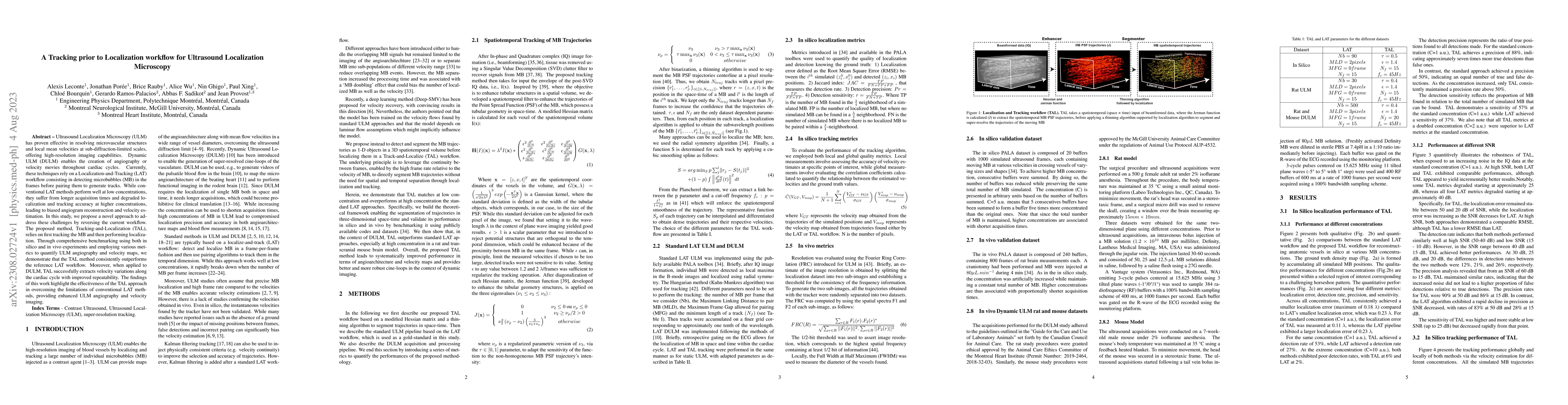
Ultrasound Localization Microscopy (ULM) has proven effective in resolving microvascular structures and local mean velocities at sub-diffraction-limited scales, offering high-resolution imaging capa...
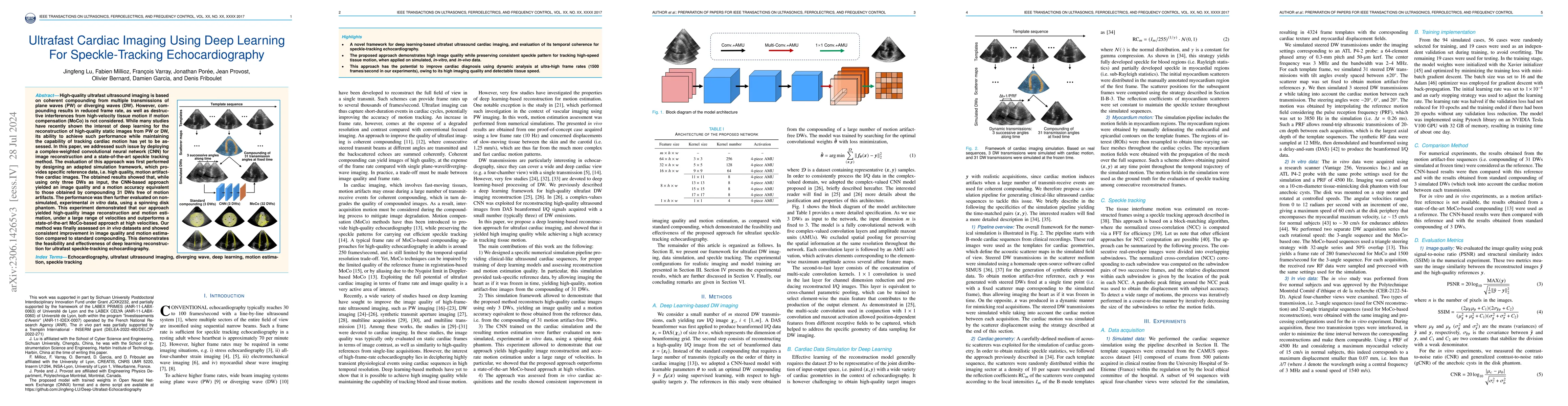
High-quality ultrafast ultrasound imaging is based on coherent compounding from multiple transmissions of plane waves (PW) or diverging waves (DW). However, compounding results in reduced frame rate...
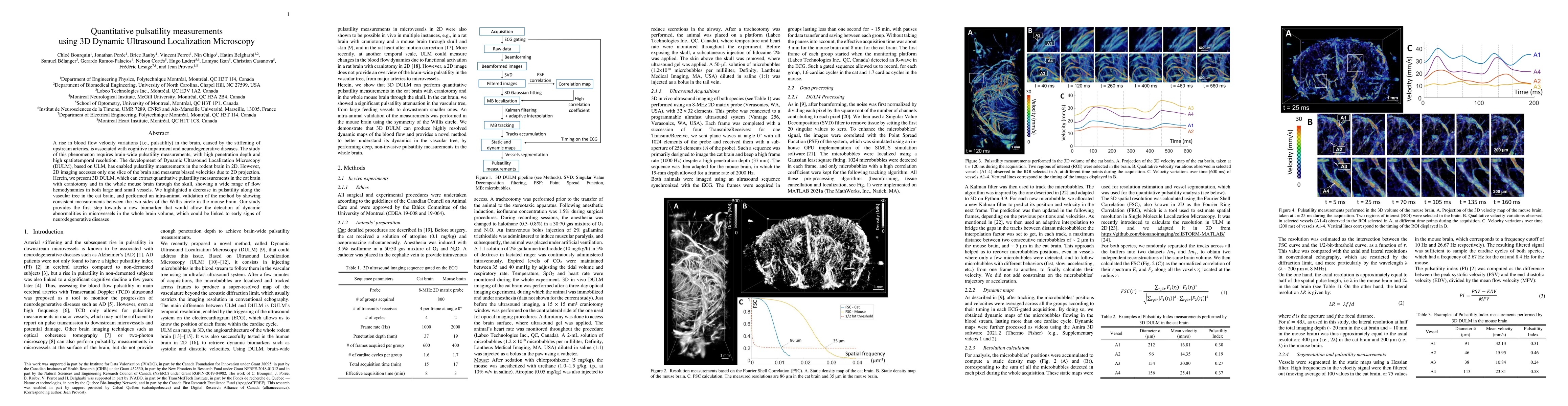
A rise in blood flow velocity variations (i.e., pulsatility) in the brain, caused by the stiffening of upstream arteries, is associated with cognitive impairment and neurodegenerative diseases. The ...
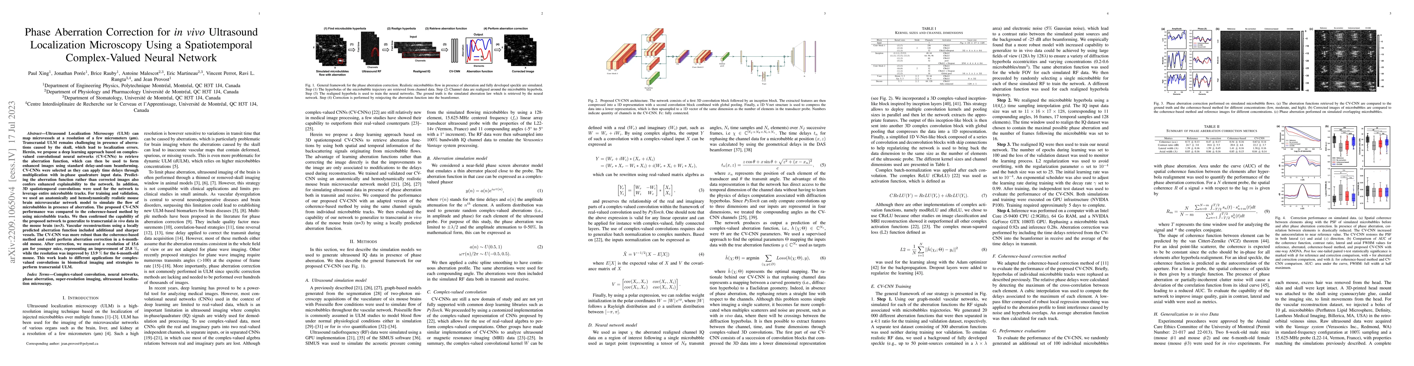
Ultrasound Localization Microscopy (ULM) can map microvessels at a resolution of a few micrometers (\mu m). Transcranial ULM remains challenging in presence of aberrations caused by the skull, which...
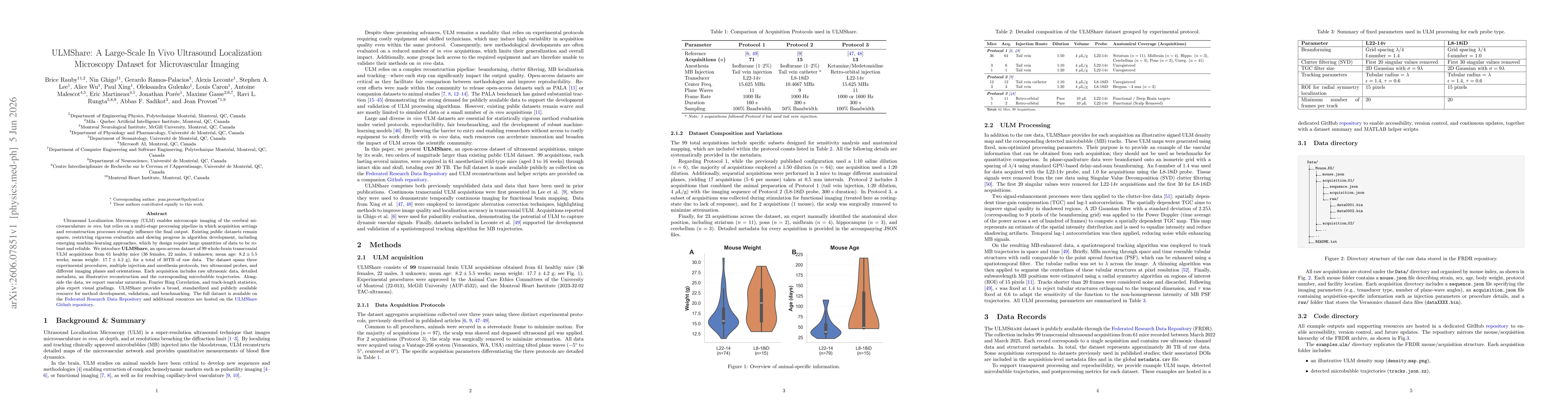
Ultrasound Localization Microscopy (ULM) enables microscopic imaging of the cerebral microvasculature in vivo, but relies on a multi-stage processing pipeline in which acquisition settings and reconst...