Academic Profile
Statistics
Similar Authors
Papers on arXiv
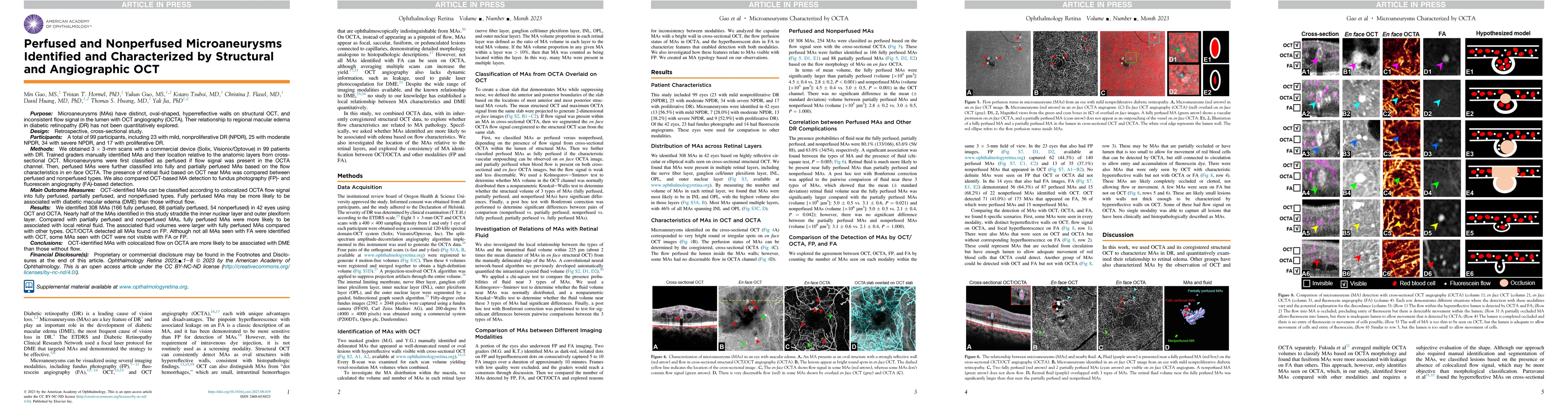
Purpose: Microaneurysms (MAs) have distinct, oval-shaped, hyperreflective walls on structural OCT, and inconsistent flow signal in the lumen with OCT angiography (OCTA). Their relationship to region...
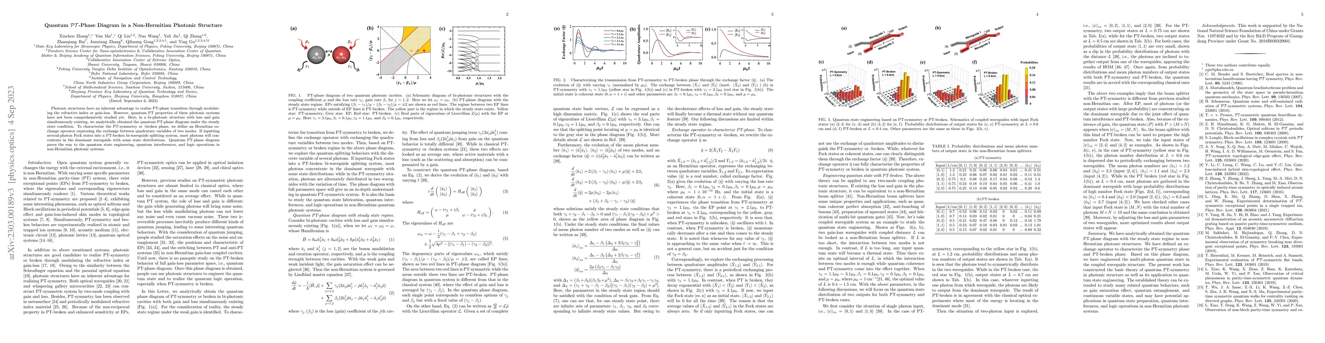
Photonic structures have an inherent advantage to realize PT-phase transition through modulating the refractive index or gain-loss. However, quantum PT properties of these photonic systems have not ...
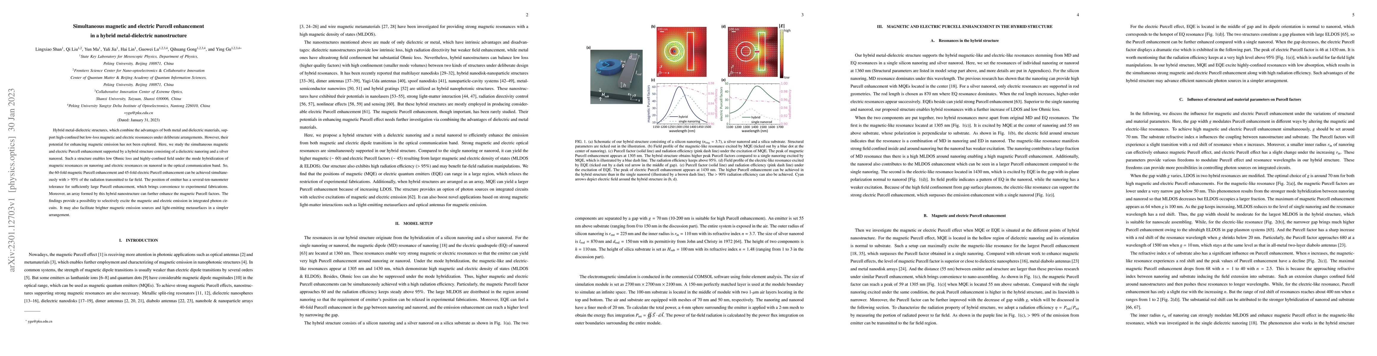
Hybrid metal-dielectric structures, which combine the advantages of both metal and dielectric materials, support high-confined but low-loss magnetic and electric resonances under deliberate arrangem...
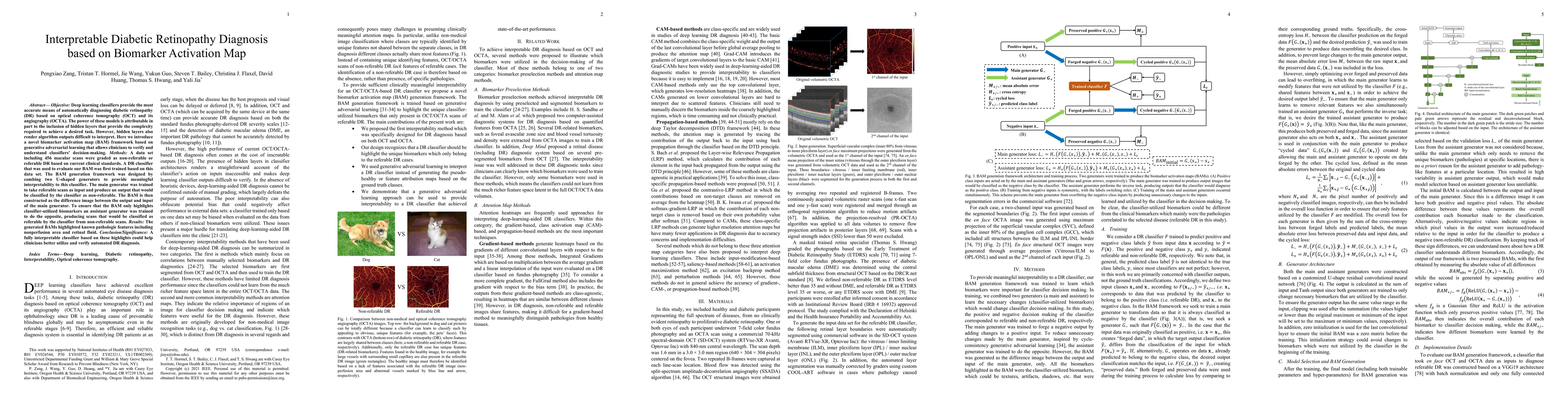
Deep learning classifiers provide the most accurate means of automatically diagnosing diabetic retinopathy (DR) based on optical coherence tomography (OCT) and its angiography (OCTA). The power of t...
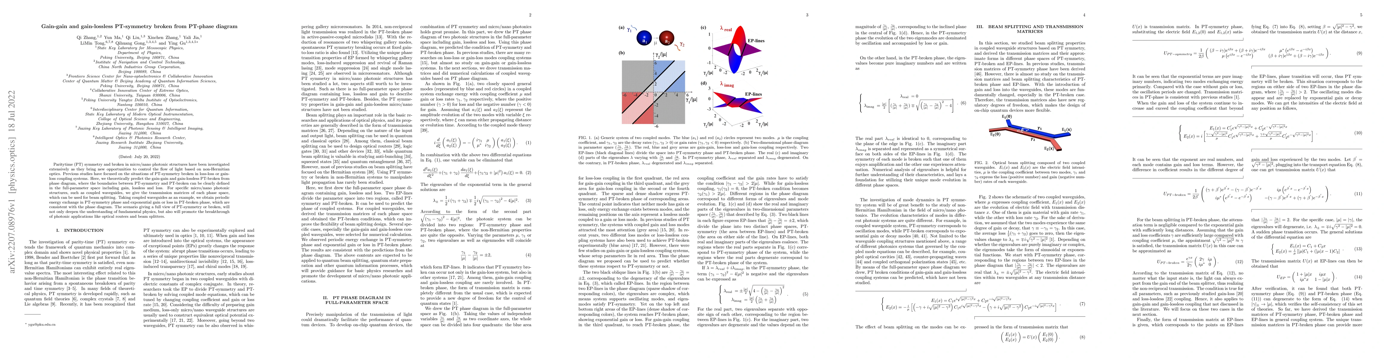
Parity-time (PT) symmetry and broken in micro/nano photonic structures have been investigated extensively as they bring new opportunities to control the flow of light based on non-Hermitian optics. ...
Purpose: To evaluate nerve fiber layer (NFL) reflectance for glaucoma diagnosis. Methods: Participants were imaged with 4.5X4.5-mm volumetric disc scans using spectral-domain optical coherence tomog...
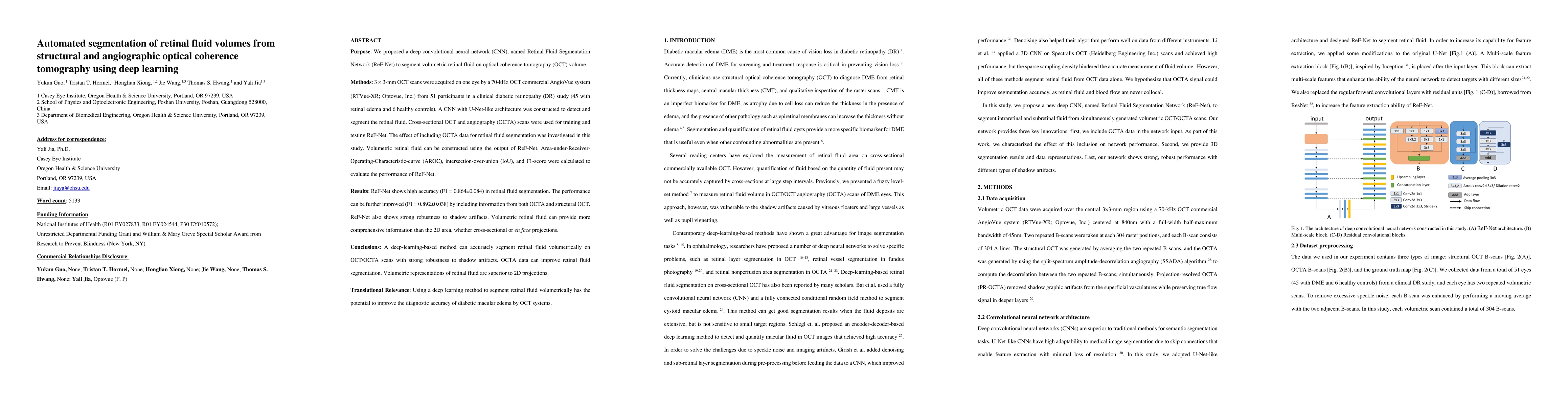
Purpose: We proposed a deep convolutional neural network (CNN), named Retinal Fluid Segmentation Network (ReF-Net) to segment volumetric retinal fluid on optical coherence tomography (OCT) volume. M...
To develop a new method to quantify nonperfused retinal capillaries (NPCs) by using co-registered optical coherence tomography (OCT) and OCT angiography (OCTA), and to evaluate NPCs in eyes with age-r...
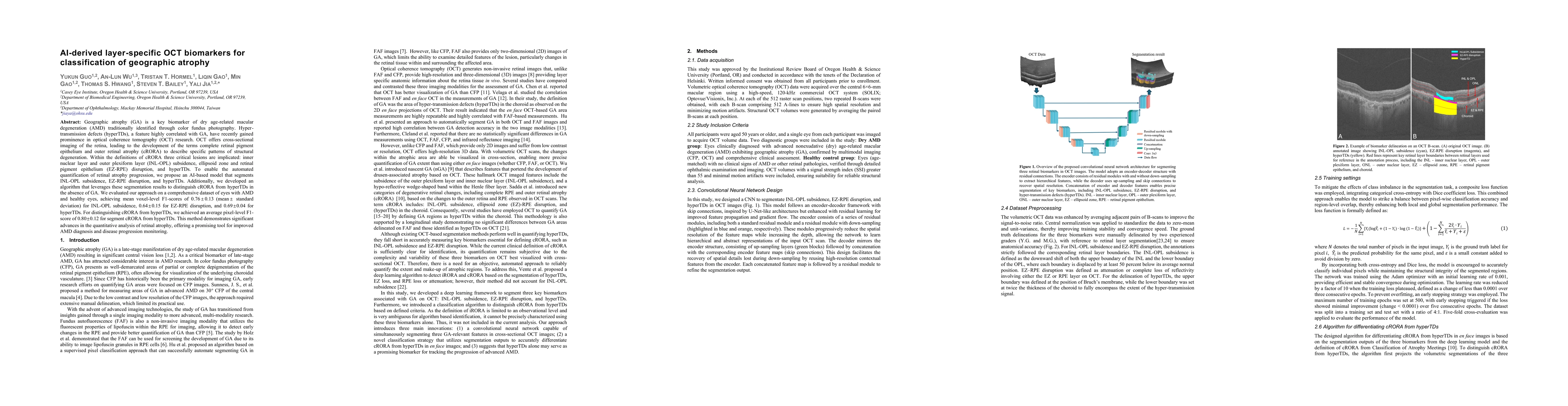
Geographic atrophy (GA) is a key biomarker of dry age-related macular degeneration (AMD) traditionally identified through color fundus photography. Hyper-transmission defects (hyperTDs), a feature hig...
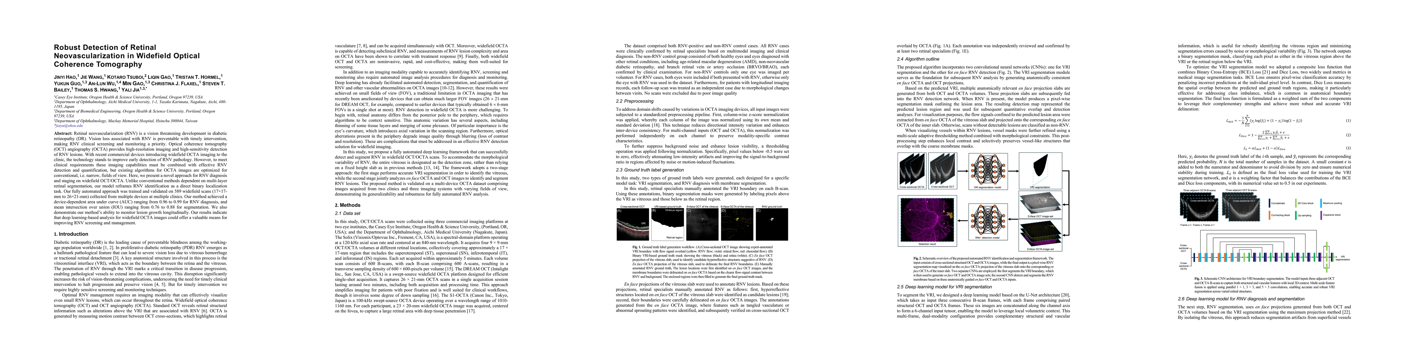
Retinal neovascularization (RNV) is a vision threatening development in diabetic retinopathy (DR). Vision loss associated with RNV is preventable with timely intervention, making RNV clinical screenin...
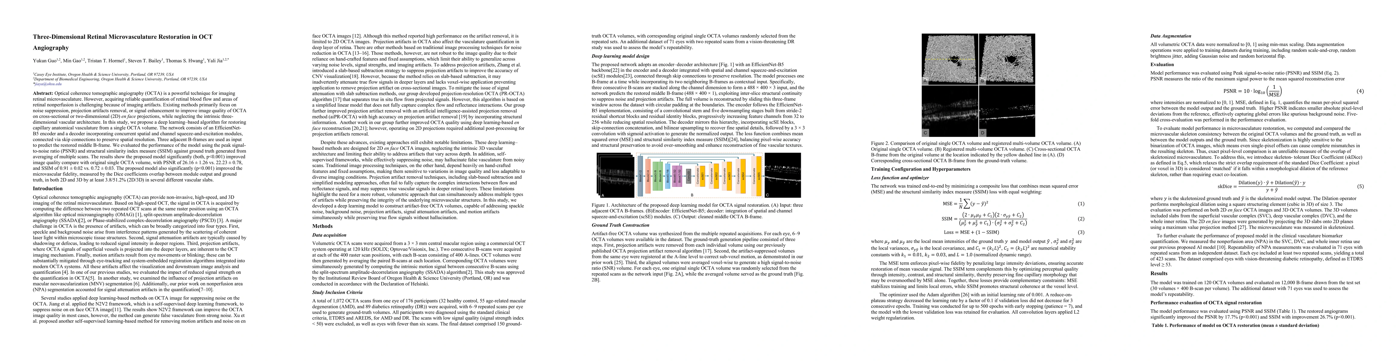
Optical coherence tomographic angiography (OCTA) is a powerful technique for imaging retinal microvasculature. However, acquiring reliable quantification of retinal blood flow and areas of retinal non...
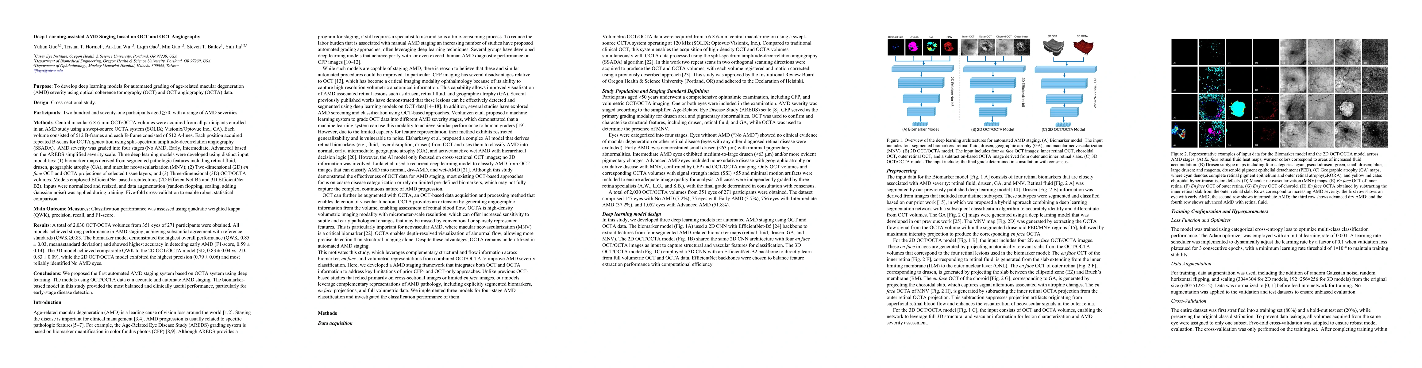
To develop and evaluate deep learning models for automated grading of age-related macular degeneration (AMD) severity using optical coherence tomography (OCT) and OCT angiography (OCTA) data. Two hund...