A deep learning pipeline using a three-frame input strategy where three adjacent B-frames are fed into an EfficientNet-B5 encoder with a decoder that incorporates concurrent spatial and channel squeeze-and-excitation modules and skip connections to preserve spatial resolution. The network predicts the restored middle B-frame from the surrounding frames, effectively denoising and reconstructing 3D capillary vasculature from a single OCTA volume. Model training leverages ground truth generated by averaging multiple scans and evaluation uses PSNR, SSIM, and Dice coefficient against ground truth across 2D and 3D vascular slabs.
Three-Dimensional Retinal Microvasculature Restoration in OCT Angiography
Publication
Metrics
AI Quick Summary
A deep learning method using an EfficientNet-B5 encoder and a skip-connected decoder restores 3D retinal capillary structure from a single OCTA volume. By inputting three adjacent frames, the model significantly improves image quality (PSNR 26.16 vs 22.23; SSIM 0.91 vs 0.72) and microvascular fidelity ( Dice gain >3.8% in 2D, >51.2% in 3D) compared with ground-truth averages.
Quick Answers
What is "Three-Dimensional Retinal Microvasculature Restoration in OCT Angiography" about?
A deep learning method using an EfficientNet-B5 encoder and a skip-connected decoder restores 3D retinal capillary structure from a single OCTA volume. By inputting three adjacent frames, the model significantly improves image quality (PSNR 26.16 vs 22.23; SSIM 0.91 vs 0.72) and microvascular fidelity ( Dice gain >3.8% in 2D, >51.2% in 3D) compared with ground-truth averages.
What methodology did the authors use?
A deep learning pipeline using a three-frame input strategy where three adjacent B-frames are fed into an EfficientNet-B5 encoder with a decoder that incorporates concurrent spatial and channel squeeze-and-excitation modules and skip connections to preserve spatial resolution. The network predicts the restored middle B-frame from the surrounding frames, effectively denoising and reconstructing 3D capillary vasculature from a single OCTA volume.... More in Methodology →
What are the key results?
PSNR improved to 26.16 ± 1.26 compared with 22.23 ± 0.78 for the original single OCTA volume. — SSIM improved to 0.91 ± 0.02 compared with 0.72 ± 0.03 for the original single OCTA volume. More in Key Results →
Why is this work significant?
Demonstrates that a volumetric, artifact-aware deep learning approach can restore 3D retinal microvasculature from a single OCTA volume, significantly enhancing image quality and structural fidelity over traditional single-volume OCTA approaches while addressing multiple artifact types concurrently. More in Significance →
What are the main limitations?
Ground truth relies on averaging multiple scans, which may not be feasible in all clinical settings due to acquisition time and patient motion. — Performance is reported on a specific OCTA system and scan protocol; generalization to other devices and settings remains to be validated. More in Limitations →
Abstract
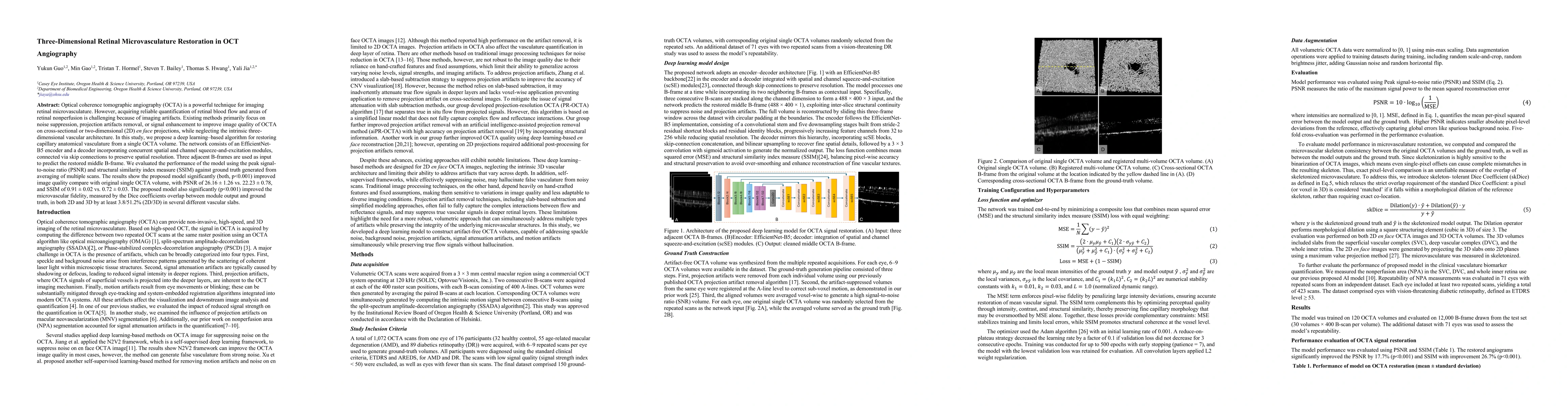
Optical coherence tomographic angiography (OCTA) is a powerful technique for imaging retinal microvasculature. However, acquiring reliable quantification of retinal blood flow and areas of retinal nonperfusion is challenging because of imaging artifacts. Existing methods primarily focus on noise suppression, projection artifact removal, or signal enhancement to improve the image quality of OCTA in cross-sectional or two-dimensional (2D) en face projections, while neglecting the intrinsic three-dimensional vascular architecture. In this study, we propose a deep learning-based algorithm for restoring capillary anatomical vasculature from a single OCTA volume. The network consists of an EfficientNet-B5 encoder and a decoder incorporating concurrent spatial and channel squeeze-and-excitation modules, connected via skip connections to preserve spatial resolution. Three adjacent B-frames are used as input to predict the restored middle B-frame. We evaluated the performance of the model using the peak signal-to-noise ratio (PSNR) and structural similarity index measure (SSIM) against ground truth generated from averaging multiple scans. The results show that the proposed model significantly (both p < 0.001) improved image quality compared with the original single OCTA volume, with a PSNR of 26.16 +/- 1.26 vs. 22.23 +/- 0.78 and an SSIM of 0.91 +/- 0.02 vs. 0.72 +/- 0.03. The proposed model also significantly (p < 0.001) improved microvascular fidelity, measured by the Dice coefficient overlap between the model output and ground truth, in both 2D and 3D by at least 3.8% and 51.2%, respectively, across several different vascular slabs.
Key Findings, in focus
Seven facets of this paper, analysed and brought into focus by AI.
Demonstrates that a volumetric, artifact-aware deep learning approach can restore 3D retinal microvasculature from a single OCTA volume, significantly enhancing image quality and structural fidelity over traditional single-volume OCTA approaches while addressing multiple artifact types concurrently.
- PSNR improved to 26.16 ± 1.26 compared with 22.23 ± 0.78 for the original single OCTA volume.
- SSIM improved to 0.91 ± 0.02 compared with 0.72 ± 0.03 for the original single OCTA volume.
- Dice coefficient overlap indicating microvascular fidelity improved by at least 3.8% in 2D and 51.2% in 3D across several vascular slabs.
Demonstrates that a volumetric, artifact-aware deep learning approach can restore 3D retinal microvasculature from a single OCTA volume, significantly enhancing image quality and structural fidelity over traditional single-volume OCTA approaches while addressing multiple artifact types concurrently.
Proposes a 3-frame input deep learning framework with EfficientNet-B5 encoding and a spatial-channel squeeze-and-excitation based decoder, linked by skip connections, to reconstruct artifact-free 3D OCTA volumes from a single input volume.
Addresses the limitation of prior methods by focusing on three-dimensional vascular restoration from a single OCTA volume rather than 2D projections or single-scan denoising, incorporating concurrent spatial and channel attention to preserve fine microvascular architecture across depth.
- Ground truth relies on averaging multiple scans, which may not be feasible in all clinical settings due to acquisition time and patient motion.
- Performance is reported on a specific OCTA system and scan protocol; generalization to other devices and settings remains to be validated.
- Evaluation focuses on PSNR/SSIM and Dice metrics; clinical impact on diagnostic accuracy for pathological nonperfusion or flow quantification needs further study.
- Extend validation to diverse OCTA devices and larger, multi-center cohorts to assess generalizability.
- Investigate real-time or near-real-time inference to enable clinical deployment without requiring multiple scans for ground truth.
- Explore integration with downstream quantitative metrics for retinal perfusion and nonperfusion area analysis to assess clinical utility.
Discussion 0