Summary
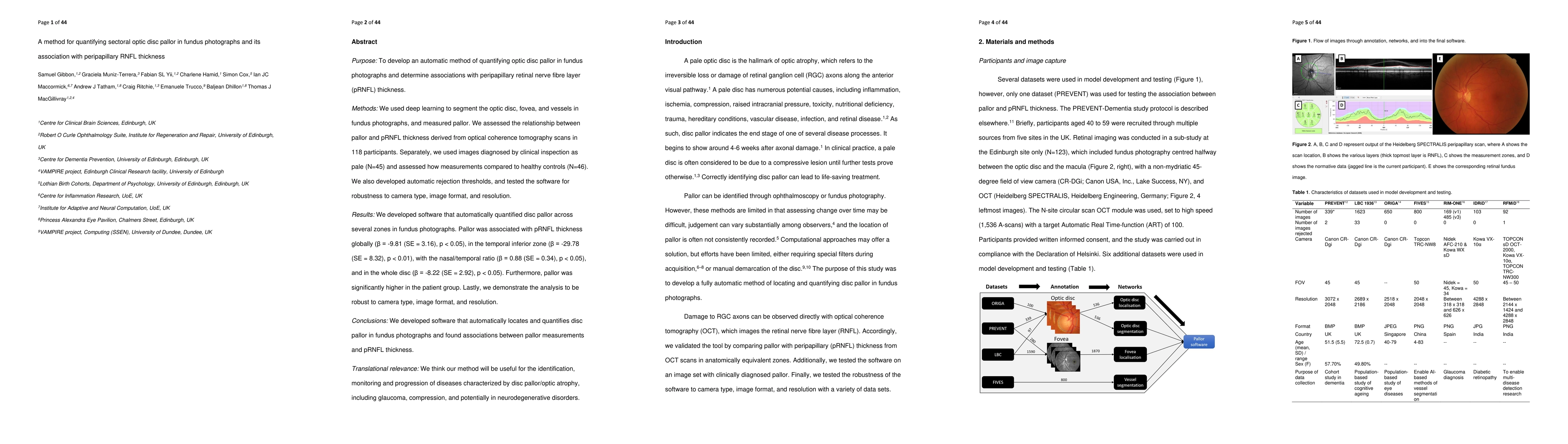
Purpose: To develop an automatic method of quantifying optic disc pallor in fundus photographs and determine associations with peripapillary retinal nerve fibre layer (pRNFL) thickness. Methods: We used deep learning to segment the optic disc, fovea, and vessels in fundus photographs, and measured pallor. We assessed the relationship between pallor and pRNFL thickness derived from optical coherence tomography scans in 118 participants. Separately, we used images diagnosed by clinical inspection as pale (N=45) and assessed how measurements compared to healthy controls (N=46). We also developed automatic rejection thresholds, and tested the software for robustness to camera type, image format, and resolution. Results: We developed software that automatically quantified disc pallor across several zones in fundus photographs. Pallor was associated with pRNFL thickness globally (\b{eta} = -9.81 (SE = 3.16), p < 0.05), in the temporal inferior zone (\b{eta} = -29.78 (SE = 8.32), p < 0.01), with the nasal/temporal ratio (\b{eta} = 0.88 (SE = 0.34), p < 0.05), and in the whole disc (\b{eta} = -8.22 (SE = 2.92), p < 0.05). Furthermore, pallor was significantly higher in the patient group. Lastly, we demonstrate the analysis to be robust to camera type, image format, and resolution. Conclusions: We developed software that automatically locates and quantifies disc pallor in fundus photographs and found associations between pallor measurements and pRNFL thickness. Translational relevance: We think our method will be useful for the identification, monitoring and progression of diseases characterized by disc pallor/optic atrophy, including glaucoma, compression, and potentially in neurodegenerative disorders.
AI Key Findings
Get AI-generated insights about this paper's methodology, results, and significance.
Paper Details
PDF Preview
Key Terms
Similar Papers
Found 4 papersPredicting Retinal Nerve Fiber Layer Thickness From Ocular Hypertension Treatment Study Optic Disc Photographs.
Liu, James C, Jammal, Alessandro A, Scherer, Rafael et al.
No citations found for this paper.
Comments (0)