This paper introduces a multiresolution convolutional neural network with a nested encoder-decoder architecture to annotate complex reflectance confocal microscopy images of skin, aiding in melanoma diagnosis. The network addresses classification challenges due to image complexity and partial labeling, achieving high sensitivity and specificity in pattern annotation.
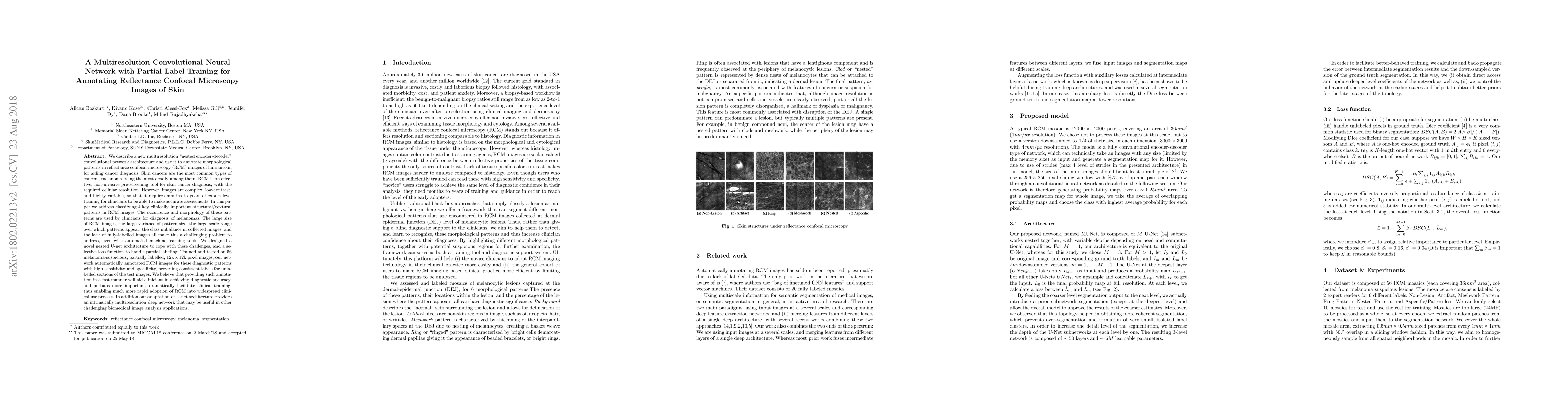
We describe a new multiresolution "nested encoder-decoder" convolutional network architecture and use it to annotate morphological patterns in reflectance confocal microscopy (RCM) images of human skin for aiding cancer diagnosis. Skin cancers are the most common types of cancers, melanoma being the deadliest among them. RCM is an effective, non-invasive pre-screening tool for skin cancer diagnosis, with the required cellular resolution. However, images are complex, low-contrast, and highly variable, so that clinicians require months to years of expert-level training to be able to make accurate assessments. In this paper, we address classifying 4 key clinically important structural/textural patterns in RCM images. The occurrence and morphology of these patterns are used by clinicians for diagnosis of melanomas. The large size of RCM images, the large variance of pattern size, the large-scale range over which patterns appear, the class imbalance in collected images, and the lack of fully-labeled images all make this a challenging problem to address, even with automated machine learning tools. We designed a novel nested U-net architecture to cope with these challenges, and a selective loss function to handle partial labeling. Trained and tested on 56 melanoma-suspicious, partially labeled, 12k x 12k pixel images, our network automatically annotated diagnostic patterns with high sensitivity and specificity, providing consistent labels for unlabeled sections of the test images. Providing such annotation will aid clinicians in achieving diagnostic accuracy, and perhaps more important, dramatically facilitate clinical training, thus enabling much more rapid adoption of RCM into widespread clinical use process. In addition, our adaptation of U-net architecture provides an intrinsically multiresolution deep network that may be useful in other challenging biomedical image analysis applications.
Get AI-generated insights about this paper's methodology, results, and significance.
Current paper (gray), citations (green), references (blue)
Chris Keum, Hong-Jun Yoon, Alexander Witkowski et al.
Xi Chen, Mikhail E. Kandel, Gabriel Popescu et al.
Title | Authors | Year | Actions |
---|---|---|---|
DermoGAN: multi-task cycle generative adversarial networks for unsupervised automatic cell identification on in-vivo reflectance confocal microscopy images of the human epidermis
|
I. Lboukili, Georgios Stamatas, Xavier Descombes
|
2024
|
View Paper |
Recent Advancements and Perspectives in the Diagnosis of Skin Diseases Using Machine Learning and Deep Learning: A Review
|
Junpeng Zhang, Fan Zhong, Kaiqiao He, Mengqi Ji, Shuli Li, Chunying Li
|
2023
|
View Paper |
Age‐dependent changes in epidermal architecture explored using an automated image analysis algorithm on in vivo reflectance confocal microscopy images
|
I. Lboukili, G. Stamatas, X. Descombes
|
2023
|
View Paper |
Automatic granular and spinous epidermal cell identification and analysis on in vivo reflectance confocal microscopy images using cell morphological features
|
I. Lboukili, G. Stamatas, X. Descombes
|
2023
|
View Paper |
Automating reflectance confocal microscopy image analysis for dermatological research: a review
|
I. Lboukili, G. Stamatas, X. Descombes
|
2022
|
View Paper |
Automatic cell identification and analysis on in vivo reflectance confocal microscopy images of the human epidermis
|
I. Lboukili, X. Descombes, G. Stamatas
|
2022
|
View Paper |
Deep learning on reflectance confocal microscopy improves Raman spectral diagnosis of basal cell carcinoma
|
Mengkun Chen, Xu Feng, Matthew C. Fox, J. Reichenberg, F. C. Lopes, Katherine R. Sebastian, M. Markey, J. Tunnell
|
2022
|
View Paper |
A State-of-the-art Survey of U-Net in Microscopic Image Analysis: from Simple Usage to Structure Mortification
|
Jian Wu, Wanli Liu, Chen Li, Tao Jiang, Islam Mohammad Shariful, Hongzan Sun, Xiaoqi Li, Xintong Li, Xinyu Huang, M. Grzegorzek
|
2022
|
View Paper |
Artificial Intelligence-Based Approaches to Reflectance Confocal Microscopy Image Analysis in Dermatology
|
A. Malciu, M. Lupu, V. Voiculescu
|
2022
|
View Paper |
Multi_Scale_Tools: A Python Library to Exploit Multi-Scale Whole Slide Images
|
Niccolò Marini, Sebastian Otálora, Damian Podareanu, Mart van Rijthoven, J. Laak, F. Ciompi, H. Müller, M. Atzori
|
2021
|
View Paper |
Deep Learning for Basal Cell Carcinoma Detection for Reflectance Confocal Microscopy.
|
Gabriele Campanella, C. Navarrete-Dechent, K. Liopyris, J. Monnier, Saud Aleissa, Brahmteg Minhas, A. Scope, C. Longo, P. Guitera, G. Pellacani, K. Kose, A. Halpern, Thomas J. Fuchs, Manu Jain
|
2021
|
View Paper |
Learning-based local quality assessment of reflectance confocal microscopy images for dermatology applications
|
Miroslawa Sikorska, A. Skalski, Marek Wodziński, A. Witkowski, G. Pellacani, J. Ludzik
|
2021
|
View Paper |
Semantic segmentation of reflectance confocal microscopy mosaics of pigmented lesions using weak labels
|
Marissa D'Alonzo, A. Bozkurt, C. Alessi-Fox, M. Gill, D. Brooks, M. Rajadhyaksha, K. Kose, Jennifer G. Dy
|
2021
|
View Paper |
A Clinical Perspective on the Automated Analysis of Reflectance Confocal Microscopy in Dermatology
|
J. Mehrabi, E. Baugh, Alexander Fast, G. Lentsch, M. Balu, Bonnie A. Lee, K. Kelly
|
2021
|
View Paper |
MSN-Net: a multi-scale context nested U-Net for liver segmentation
|
Tongle Fan, Guanglei Wang, Xia Wang, Yan Li, Hongrui Wang
|
2021
|
View Paper |
Automatic Quality Assessment of Reflectance Confocal Microscopy Mosaics using Attention-Based Deep Neural Network
|
Marek Wodziński, Miroslawa Pajak, A. Skalski, A. Witkowski, G. Pellacani, J. Ludzik
|
2020
|
View Paper |
Speckle-free, near-infrared portable confocal microscope.
|
Cheng Gong, Delaney B Stratton, C. Curiel-Lewandrowski, Dongkyun Kang
|
2020
|
View Paper |
Mu-net: Multi-scale U-net for two-photon microscopy image denoising and restoration
|
Sehyung Lee, Makiko Negishi, H. Urakubo, H. Kasai, Shin Ishii
|
2020
|
View Paper |
Segmentation of Cellular Patterns in Confocal Images of Melanocytic Lesions in vivo via a Multiscale Encoder-Decoder Network (MED-Net)
|
Kivanc Kose, A. Bozkurt, C. Alessi-Fox, M. Gill, C. Longo, G. Pellacani, J. Dy, D. Brooks, M. Rajadhyaksha
|
2020
|
View Paper |
Utilizing Machine Learning for Image Quality Assessment for Reflectance Confocal Microscopy.
|
K. Kose, A. Bozkurt, C. Fox, D. Brooks, Jennifer G. Dy, M. Rajadhyaksha, M. Gill
|
2019
|
View Paper |
Artificial Intelligence in Skin Cancer
|
O. Reiter, V. Rotemberg, K. Kose, A. Halpern
|
2019
|
View Paper |
Convolutional Neural Network Approach to Classify Skin Lesions Using Reflectance Confocal Microscopy
|
Marek Wodziński, A. Skalski, A. Witkowski, G. Pellacani, J. Ludzik
|
2019
|
View Paper |
Multi-Scale Task Multiple Instance Learning for the Classification of Digital Pathology Images with Global Annotations
|
Niccolò Marini, Sebastian Otálora, F. Ciompi, G. Silvello, S. Marchesin, S. Vatrano, Gravina Caltagirone, M. Atzori, H. Müller
|
2021
|
View Paper |
Supervised CNN Strategies for Optical Image Segmentation and Classification in Interventional Medicine
|
S. Moccia, L. Romeo, Lucia Migliorelli, E. Frontoni, P. Zingaretti
|
2020
|
View Paper |
LOW-COST, NEAR-INFRARED, SCAN-LESS PORTABLE CONFOCAL MICROSCOPE
|
Cheng Gong, Dr. Rongguang Liang, Dr. Jennifer Kehlet Barton, Christopher David Nguyen
|
View Paper |
Comments (0)