Publication
Metrics
AI Quick Summary
This paper proposes a framework for automatic ischemic stroke lesion segmentation using synthesized pseudo Diffusion-Weighted Imaging (DWI) from Computed Tomography Perfusion (CTP) images, employing Convolutional Neural Networks (CNNs) and attention-based deep learning. The framework achieved top performance in the ISLES 2018 challenge, demonstrating improved lesion segmentation accuracy compared to direct CTP-based methods.
Paper Preview
Abstract
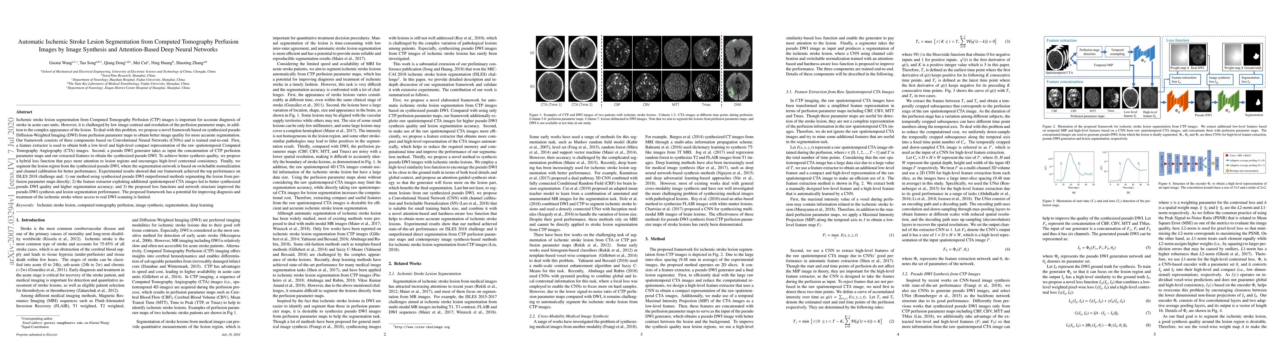
Ischemic stroke lesion segmentation from Computed Tomography Perfusion (CTP) images is important for accurate diagnosis of stroke in acute care units. However, it is challenged by low image contrast and resolution of the perfusion parameter maps, in addition to the complex appearance of the lesion. To deal with this problem, we propose a novel framework based on synthesized pseudo Diffusion-Weighted Imaging (DWI) from perfusion parameter maps to obtain better image quality for more accurate segmentation. Our framework consists of three components based on Convolutional Neural Networks (CNNs) and is trained end-to-end. First, a feature extractor is used to obtain both a low-level and high-level compact representation of the raw spatiotemporal Computed Tomography Angiography (CTA) images. Second, a pseudo DWI generator takes as input the concatenation of CTP perfusion parameter maps and our extracted features to obtain the synthesized pseudo DWI. To achieve better synthesis quality, we propose a hybrid loss function that pays more attention to lesion regions and encourages high-level contextual consistency. Finally, we segment the lesion region from the synthesized pseudo DWI, where the segmentation network is based on switchable normalization and channel calibration for better performance. Experimental results showed that our framework achieved the top performance on ISLES 2018 challenge and: 1) our method using synthesized pseudo DWI outperformed methods segmenting the lesion from perfusion parameter maps directly; 2) the feature extractor exploiting additional spatiotemporal CTA images led to better synthesized pseudo DWI quality and higher segmentation accuracy; and 3) the proposed loss functions and network structure improved the pseudo DWI synthesis and lesion segmentation performance.
AI Key Findings
Get AI-generated insights about this paper's methodology, results, significance, and more — seven facets brought into focus.
Impact
Paper Details
Authors
PDF Preview
Key Terms
Citation Network
Current paper (gray), citations (green), references (blue)
Display is limited for performance on very large graphs.
Discussion 0