A Python package called darfix provides tools for analyzing dark-field X-ray microscopy data, including automatic extraction of instrument angle settings and support for processing large image sets with limited memory resources.
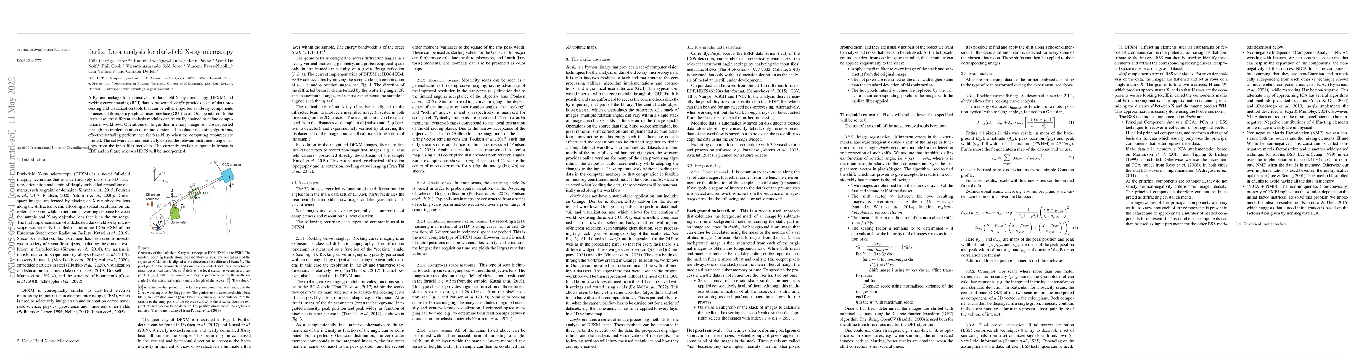
A Python package for the analysis of dark-field X-ray microscopy (DFXM) and rocking curve imaging (RCI) data is presented. \textit{darfix} provides a set of data processing and visualization tools that can be either imported as library components or accessed through a graphical user interface (GUI) as an Orange add-on. In the latter case, the different analysis modules can be easily chained to define computational workflows. Operations on larger-than-memory image sets are supported through the implementation of online versions of the data processing algorithms, effectively trading performance for feasibility when the computing resources are limited. The software can automatically extract the relevant instrument angle settings from the input files metadata. The currently available input file format is EDF and in future releases HDF5 will be incorporated.
Get AI-generated insights about this paper's methodology, results, and significance.
Current paper (gray), citations (green), references (blue)
Henning Friis Poulsen, Sina Borgi, Grethe Winther et al.
Kaye S. Morgan, James A. Pollock, Linda C. P. Croton et al.
Henning Friis Poulsen, Can Yildirim, Carsten Detlefs et al.
K. Bhattacharya, C. K. Cocke, A. Akerson et al.
Bastian Pfau, Stefan Eisebitt, Dirk Grundler et al.
Title | Authors | Year | Actions |
---|---|---|---|
Pink-Beam Dark Field X-ray Microscopy: Expanding 3D/4D Imaging for Complex and Deformed Microstructures
|
C. Yildirim, Aditya Shukla, Yubin Zhang, Nikolas Mavrikakis, Louis Lesage, Virginia Sanna, M. Sarkis, Yaozhu Li, Michela La Bella, Carsten Detlefs, Henning F. Poulsen
|
2025
|
View Paper |
Phase-separated charge order and twinning across length scales in
<mml:math xmlns:mml="http://www.w3.org/1998/Math/MathML"><mml:mrow><mml:msub><mml:mi>CsV</mml:mi><mml:mn>3</mml:mn></mml:msub><mml:msub><mml:mi>Sb</mml:mi><mml:mn>5</mml:mn></mml:msub></mml:mrow></mml:math>
|
Jayden Plumb, A. C. Salinas, Krishnanand Mallayya, Elliot Kisiel, F. B. Carneiro, Reina Gomez, G. Pokharel, Eun-Ah Kim, Suchismita Sarker, Z. Islam, Samantha Daly, Stephen D. Wilson
|
2024
|
View Paper |
Observing formation and evolution of dislocation cells during plastic deformation
|
A. Zelenika, A. A. W. Cretton, Felix Frankus, S. Borgi, F. Grumsen, C. Yildirim, C. Detlefs, G. Winther, H. F. Poulsen
|
2024
|
View Paper |
Heterogeneous Antiferroelectric Ordering in NaNbO3–SrSnO3 Ceramics Revealed by Direct Superstructure Imaging
|
Leonardo Oliveira, Mao‐Hua Zhang, J. Koruza, Raquel Rodiquez-Lamas, Can Yildirim, Hugh Simons
|
2024
|
View Paper |
High-Resolution Full-field Structural Microscopy of the Voltage Induced Filament Formation in Neuromorphic Devices
|
Elliot Kisiel, Pavel Salev, Ishwor Poudyal, Fellipe Baptista, Fanny Rodolakis, Zhan Zhang, O. Shpyrko, Ivan K. Schuller, Z. Islam, A. Frano
|
2023
|
View Paper |
3D microstructural and strain evolution during the early stages of tensile deformation
|
A. Zelenika, C. Yildirim, C. Detlefs, R. Rodríguez-Lamas, F. Grumsen, H. Poulsen, Grethe Winther European Synchrotron Radiation Facility, D. Physics, T. Denmark, Department of Civil, M. Engineering
|
2023
|
View Paper |
High-resolution 3D strain and orientation mapping within a grain of a directed energy deposition laser additively manufactured superalloy
|
Y. Chen, Y. Tang, D. Collins, S. Clark, W. Ludwig, R. Rodríguez-Lamas, C. Detlefs, R. Reed, P. D. Lee, P. Withers, C. Yildirim
|
2023
|
View Paper |
Automatic Determination of the Weak-Beam Condition in Dark Field X-ray Microscopy
|
P. Huang, R. Coffee, L. Dresselhaus-Marais
|
2022
|
View Paper |
Simultaneous bright- and dark-field X-ray microscopy at X-ray free electron lasers
|
L. Dresselhaus-Marais, B. Kozioziemski, T. S. Holstad, T. Ræder, M. Seaberg, D. Nam, Sangsoo Kim, Sean Breckling, Sungwook Choi, M. Chollet, P. Cook, E. Folsom, E. Galtier, Arnulfo Gonzalez, T. Gorkhover, S. Guillet, Kristoffer Haldrup, M. Howard, K. Katagiri, Seonghan Kim, Sunam Kim, Sungwon Kim, Hyunjung Kim, E. Knudsen, S. Kuschel, Hae Ja Lee, Chuanlong Lin, R. McWilliams, B. Nagler, M. Nielsen, N. Ozaki, Dayeeta Pal, Ricardo Pablo Pedro, A. Saunders, F. Schoofs, T. Sekine, H. Simons, Tim Brandt van Driel, Bihan Wang, Wenge Yang, C. Yildirim, H. Poulsen, J. Eggert
|
2022
|
View Paper |
Extensive 3D mapping of dislocation structures in bulk aluminum
|
C. Yildirim, H. Poulsen, G. Winther, C. Detlefs, P. Huang, L. Dresselhaus-Marais
|
2022
|
View Paper |
Automating Dislocation Characterization in 3D Dark Field X-ray Microscopy
|
Pin-Hua Huang, Ryan Coffee, L. Dresselhaus-Marais
|
2022
|
View Paper |
Simultaneous Dark- and Bright-Field X-ray Microscopy at X-ray Free Electron Lasers
|
L. Dresselhaus-Marais, B. Kozioziemski, T. S. Holstad, Trygve, Magnus Ræder, M. Seaberg, D. Nam, Sangsoo Kim, Sean Breckling, M. Chollet, P. Cook, E. Folsom, E. Galtier, L. Gavilan, Arnulfo Gonzalez, Tais Gorhover, S. Guillet, Kristoffer Haldrup, Marylesa, Howard, K. Katagiri, Seonghan Kim, Sunam Kim, Sungwon Kim, Hyunjung, Kim, E. Knudsen, S. Kuschel, H. Lee, Chuanlong Lin, R., S. Mcwilliams, B. Nagler, N. Ozaki, Dayeeta Pal, Ricardo Pablo, Pedro, M. Nielsen, A. Saunders, F. Schoofs, Toshimori, Sekine, H. Simons, T. V. Driel, Bihan Wang, Wenge Yang, C. Yildirim, H. Poulsen, J. Eggert
|
2022
|
View Paper |
Comments (0)