Summary
A prospective study was performed on neurosurgical samples from 18 patients to evaluate the use of Full-Field Optical Coherence Tomography (FF-OCT) in brain tumor diagnosis. FF-OCT captures en face slices of tissue samples at 1\mum resolution in 3D with a typical 200\mum imaging depth. A 1cm2 specimen is scanned at a single depth and processed in about 5 minutes. This rapid imaging process is non-invasive and 30 requires neither contrast agent injection nor tissue preparation, which makes it particularly well suited to medical imaging applications. Temporal chronic epileptic parenchyma and brain tumors such as meningiomas, low- grade and high-grade gliomas, and choroid plexus papilloma were imaged. A subpopulation of neurons, myelin fibers and CNS vasculature were clearly identified. Cortex could be discriminated from white matter, but individual glial cells as astrocytes (normal or reactive) or oligodendrocytes were not observable. This study reports for the first time on the feasibility of using FF-OCT in a real-time manner as a label-free non-invasive imaging technique in an intra-operative neurosurgical clinical setting to assess tumorous glial and epileptic margins.
AI Key Findings
Get AI-generated insights about this paper's methodology, results, and significance.
Paper Details
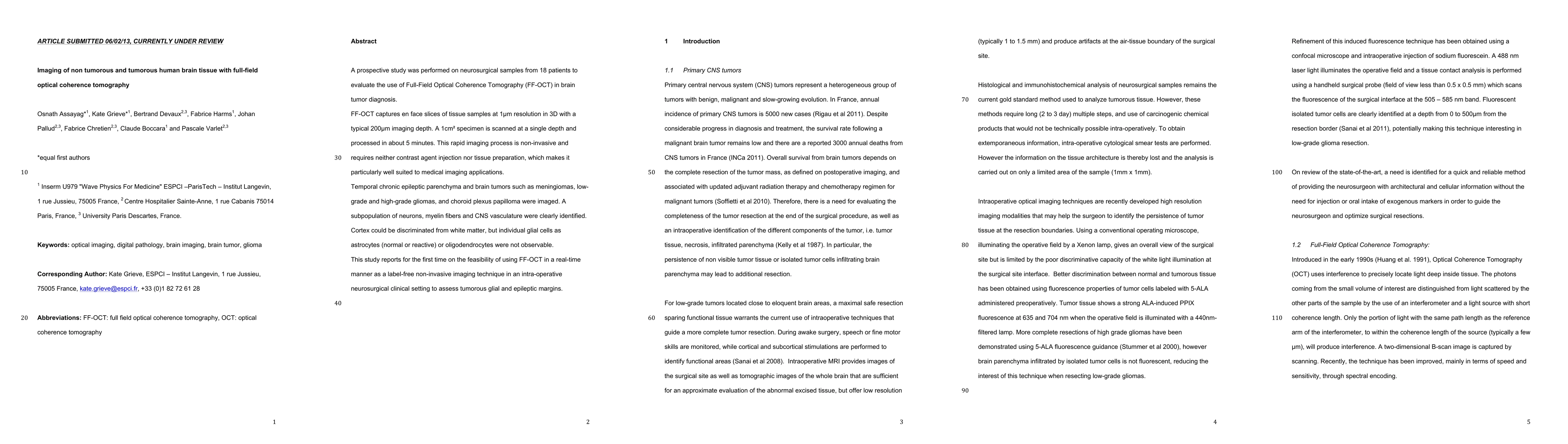
PDF Preview
Key Terms
Citation Network
Current paper (gray), citations (green), references (blue)
Display is limited for performance on very large graphs.
Similar Papers
Found 4 papersMethionine PET Findings in the Diagnosis of Brain Tumors and Non-Tumorous Mass Lesions: A Single-Center Report on 426 Cases.
Yamamoto, Ryo, Ohka, Fumiharu, Maeda, Sachi et al.
Bond-Selective Full-Field Optical Coherence Tomography
Jian Zhao, Zian Wang, Fukai Chen et al.
| Title | Authors | Year | Actions |
|---|
Comments (0)