Summary
High-resolution optical imaging methods, such as confocal microscopy and full-field optical coherence tomography, capture flat optical sections of the sample. If the sample is curved, the optical field sections through several sample layers and the view of each layer is reduced. Here we present curved-field optical coherence tomography, capable of capturing optical sections of arbitrary curvature. We test the device on a challenging task of imaging the human cornea in vivo and achieve 10x larger viewing area comparing to the clinical state-of-the-art. This enables more precise cell and nerve counts, opening a path to improved diagnosis of corneal and general health conditions (e.g. diabetes). The method is non-contact, compact and works in a single-shot, making it readily available for use in optical research and clinical practice.
AI Key Findings
Get AI-generated insights about this paper's methodology, results, and significance.
Paper Details
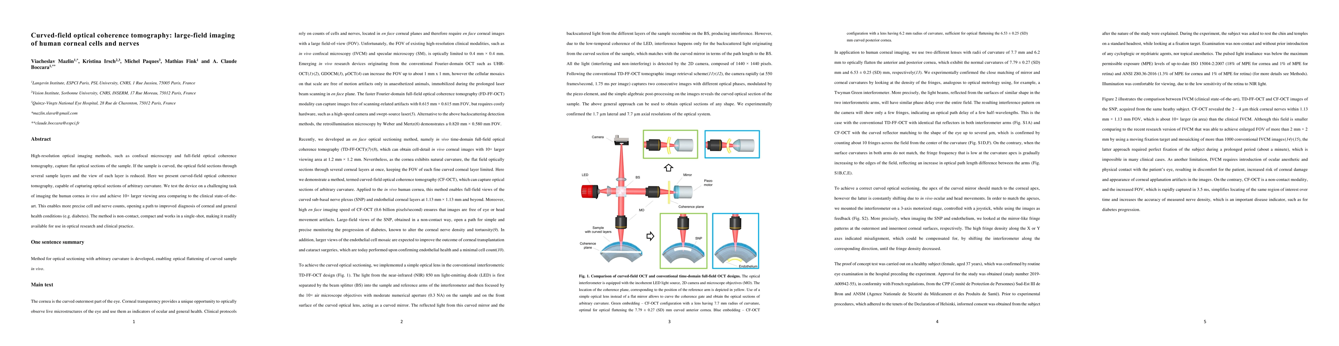
PDF Preview
Key Terms
Citation Network
Current paper (gray), citations (green), references (blue)
Display is limited for performance on very large graphs.
Similar Papers
Found 4 papers| Title | Authors | Year | Actions |
|---|
Comments (0)