Longitudinal Assessment of Lung Lesion Burden in CT

Source: ArXiv

AI Quick Summary

A new approach for automatically segmenting lung lesions and tracking total tumor burden over time was developed, outperforming a model with anatomical priors in terms of precision and sensitivity.

Share: Share on X LinkedIn

Summary

In the U.S., lung cancer is the second major cause of death. Early detection of suspicious lung nodules is crucial for patient treatment planning, management, and improving outcomes. Many approaches for lung nodule segmentation and volumetric analysis have been proposed, but few have looked at longitudinal changes in total lung tumor burden. In this work, we trained two 
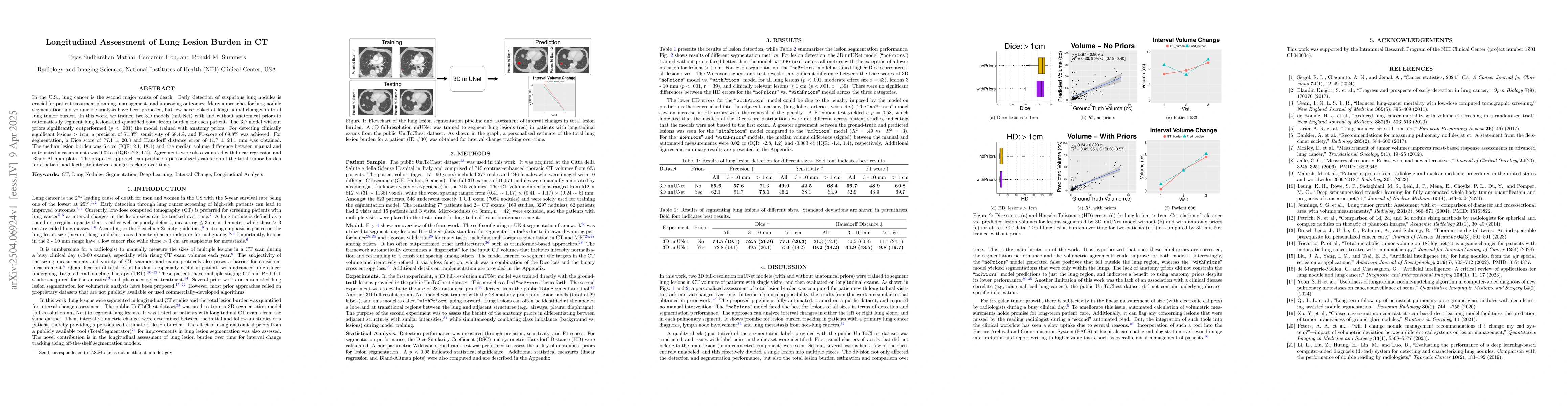
3D models (nnUNet) with and without anatomical priors to automatically segment lung lesions and quantified total lesion burden for each patient. The 3D model without priors significantly outperformed ($p < .001$) the model trained with anatomy priors. For detecting clinically significant lesions $>$ 1cm, a precision of 71.3\%, sensitivity of 68.4\%, and F1-score of 69.8\% was achieved. For segmentation, a Dice score of 77.1 $\pm$ 20.3 and Hausdorff distance error of 11.7 $\pm$ 24.1 mm was obtained. The median lesion burden was 6.4 cc (IQR: 2.1, 18.1) and the median volume difference between manual and automated measurements was 0.02 cc (IQR: -2.8, 1.2). Agreements were also evaluated with linear regression and Bland-Altman plots. The proposed approach can produce a personalized evaluation of the total tumor burden for a patient and facilitate interval change tracking over time.

AI Key Findings

Generated Jun 09, 2025

Methodology

This research trained two 3D models (nnUNet) for automatic lung lesion segmentation, one with anatomical priors and one without, to quantify total lesion burden in CT scans for longitudinal assessment of lung cancer.

Key Results

  • The model without anatomical priors significantly outperformed the model with priors ($p < .001$).
  • For detecting clinically significant lesions > 1cm, precision of 71.3%, sensitivity of 68.4%, and F1-score of 69.8% was achieved.
  • Segmentation yielded a Dice score of 77.1 ± 20.3 and Hausdorff distance error of 11.7 ± 24.1 mm.
  • The median lesion burden was 6.4 cc (IQR: 2.1, 18.1) with a median volume difference of 0.02 cc (IQR: -2.8, 1.2) between manual and automated measurements.
  • The study proposed an approach for personalized evaluation of total tumor burden and interval change tracking over time.

Significance

This research is crucial for early detection and management of lung cancer, as it presents an automated method for quantifying lung lesion burden, which can aid in treatment planning and improving patient outcomes.

Technical Contribution

The development and comparison of two 3D nnUNet models for lung lesion segmentation with and without anatomical priors, demonstrating the utility of these models for longitudinal assessment of lung tumor burden.

Novelty

This work stands out by focusing on longitudinal changes in total lung tumor burden, employing nnUNet models for segmentation, and evaluating their performance against manual measurements, providing a personalized evaluation tool for patients.

Limitations

  • The study lacked association with a clinical disease correlate, such as non-small cell lung cancer, due to the public UniToChest dataset not containing underlying disease-specific information.
  • There was subjectivity in the linear measurement of tumor size by radiologists during busy clinical days.

Future Work

  • Correcting label errors in the dataset could potentially improve segmentation performance and volumetric agreements for both models.
  • Integration of automated volumetric measurement tools into clinical workflows, such as Picture Archival and Communication Systems (PACS), could enable radiologists to focus on higher-value tasks like overall clinical management of patients.

Paper Details

PDF Preview

Download PDF

Citation Network

Current paper (gray), citations (green), references (blue)

Display is limited for performance on very large graphs.

Current Paper
Citations
References
Click to view

No citations found for this paper.

Comments (0)

This website uses cookies to ensure you get the best experience. Learn more