Academic Profile
Statistics
Similar Authors
Papers on arXiv
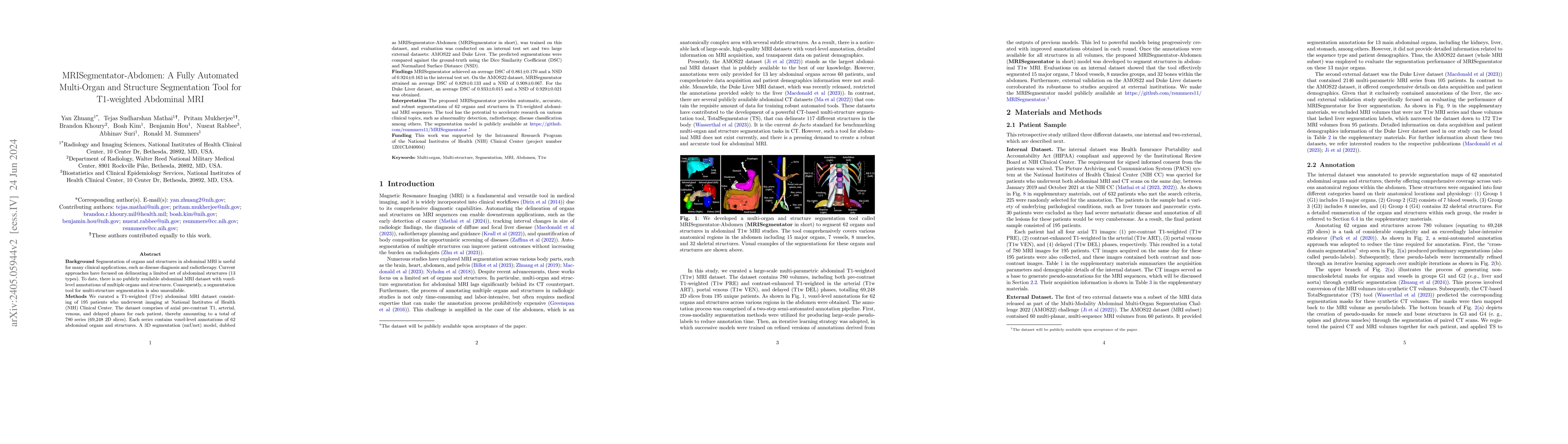
Background: Segmentation of organs and structures in abdominal MRI is useful for many clinical applications, such as disease diagnosis and radiotherapy. Current approaches have focused on delineating ...
Purpose: To evaluate the performance of an automated deep learning method in detecting ascites and subsequently quantifying its volume in patients with liver cirrhosis and ovarian cancer. Materials ...
Automatically interpreting CT scans can ease the workload of radiologists. However, this is challenging mainly due to the scarcity of adequate datasets and reference standards for evaluation. This stu...
In this paper, we introduce DRR-RATE, a large-scale synthetic chest X-ray dataset derived from the recently released CT-RATE dataset. DRR-RATE comprises of 50,188 frontal Digitally Reconstructed Rad...
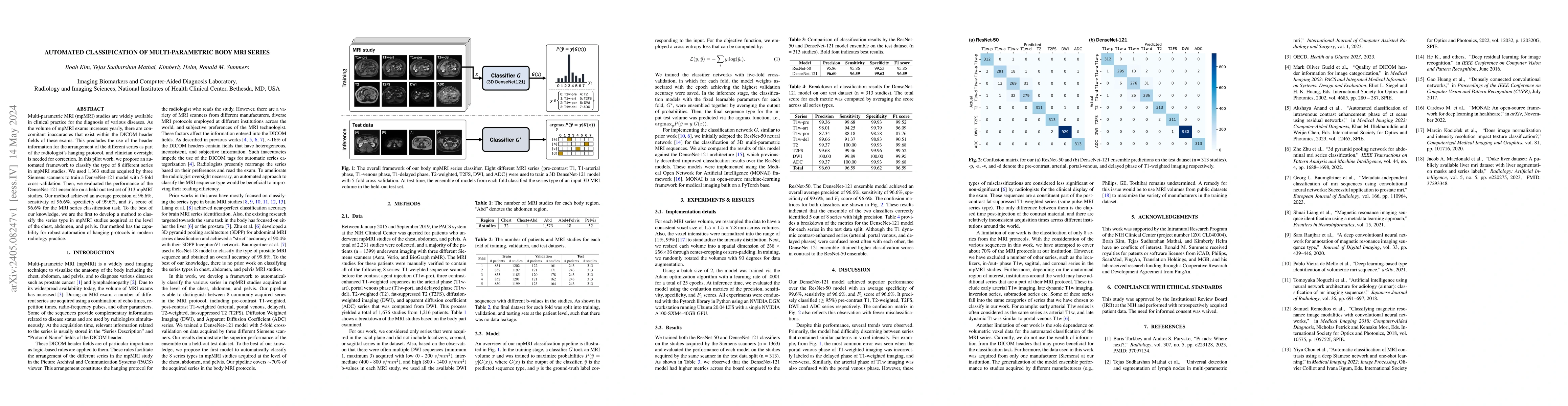
Multi-parametric MRI (mpMRI) studies are widely available in clinical practice for the diagnosis of various diseases. As the volume of mpMRI exams increases yearly, there are concomitant inaccuracie...
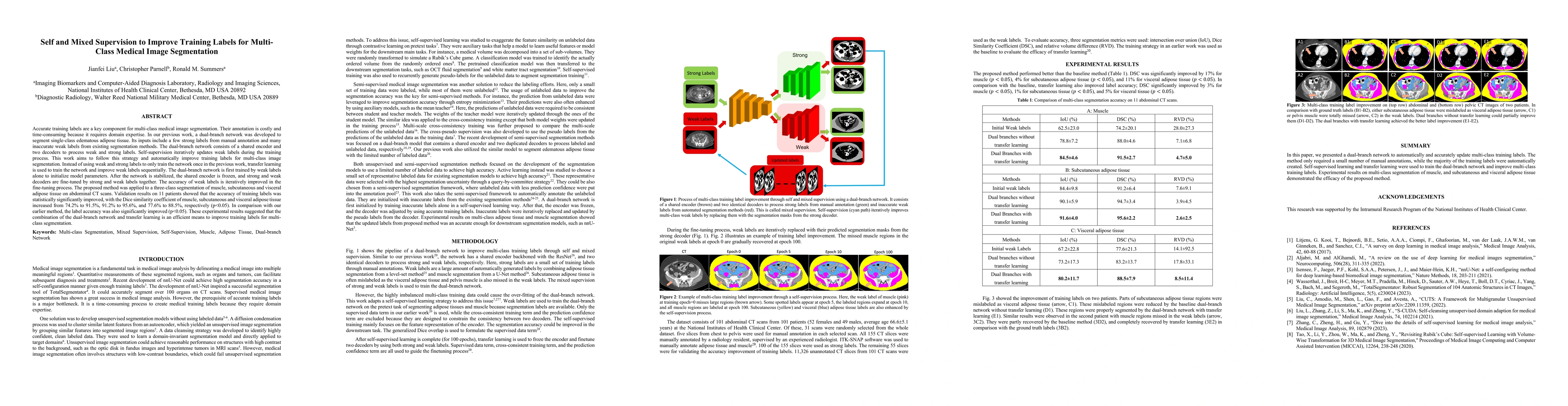
Accurate training labels are a key component for multi-class medical image segmentation. Their annotation is costly and time-consuming because it requires domain expertise. This work aims to develop...
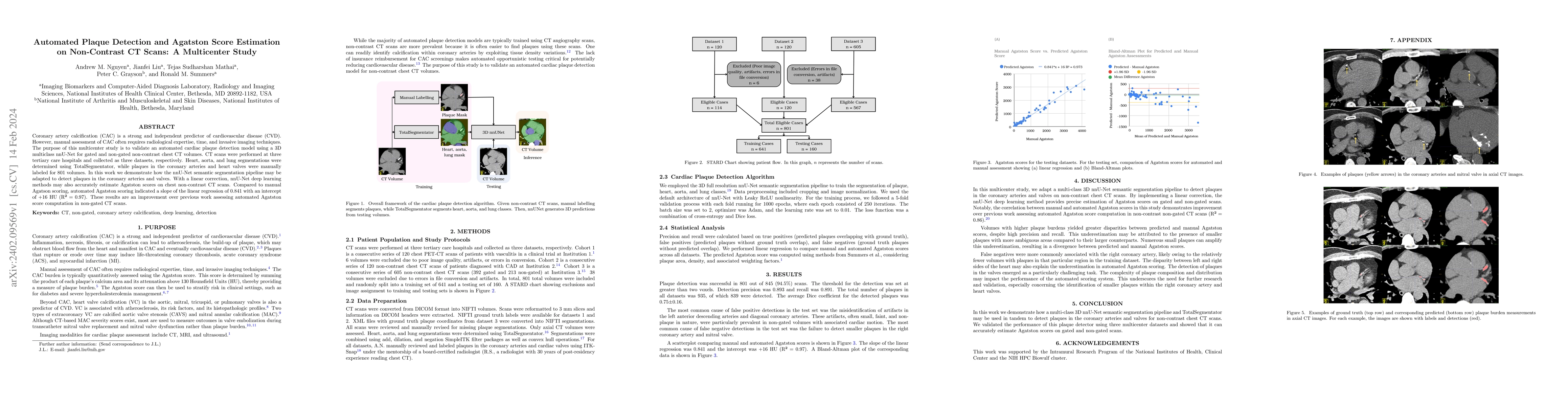
Coronary artery calcification (CAC) is a strong and independent predictor of cardiovascular disease (CVD). However, manual assessment of CAC often requires radiological expertise, time, and invasive...
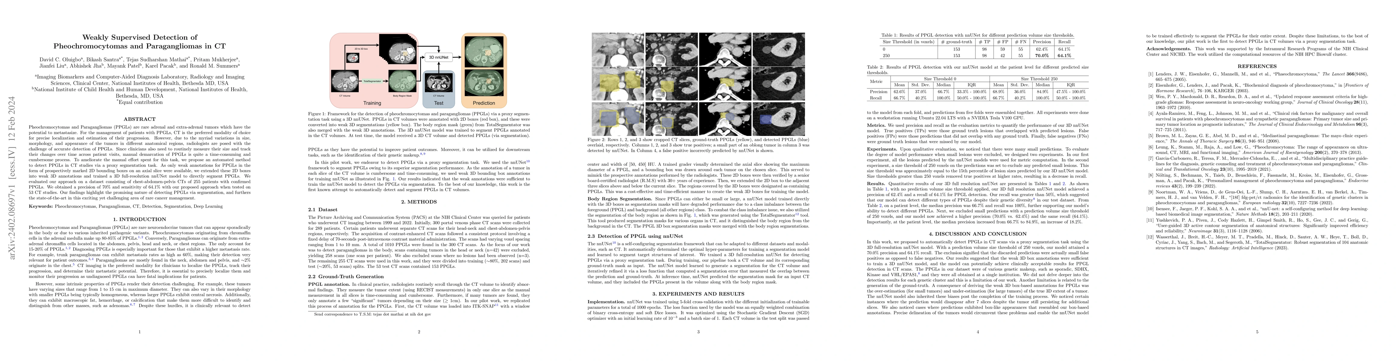
Pheochromocytomas and Paragangliomas (PPGLs) are rare adrenal and extra-adrenal tumors which have the potential to metastasize. For the management of patients with PPGLs, CT is the preferred modalit...
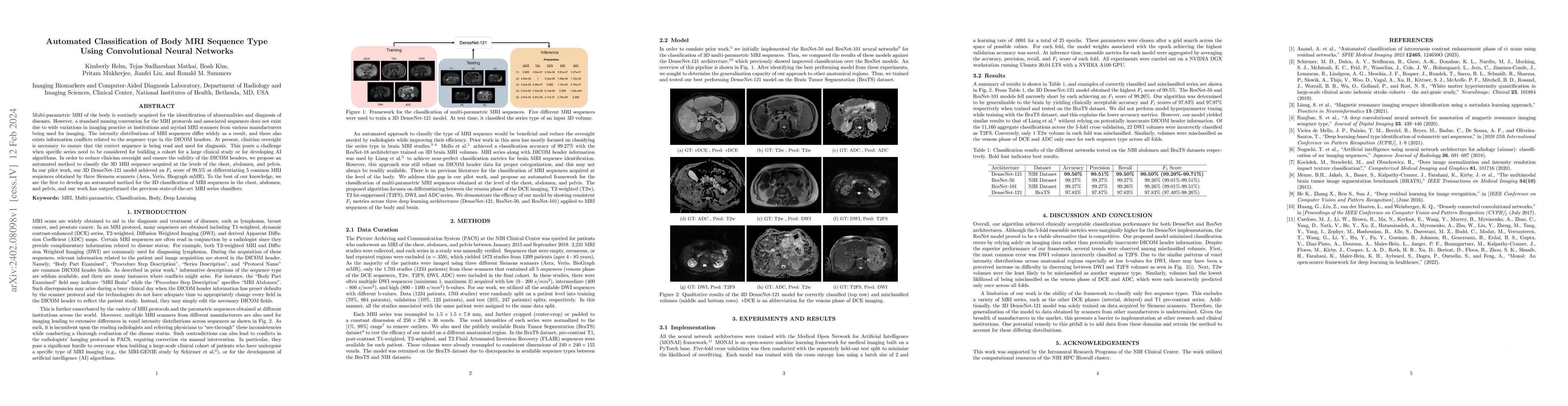
Multi-parametric MRI of the body is routinely acquired for the identification of abnormalities and diagnosis of diseases. However, a standard naming convention for the MRI protocols and associated s...
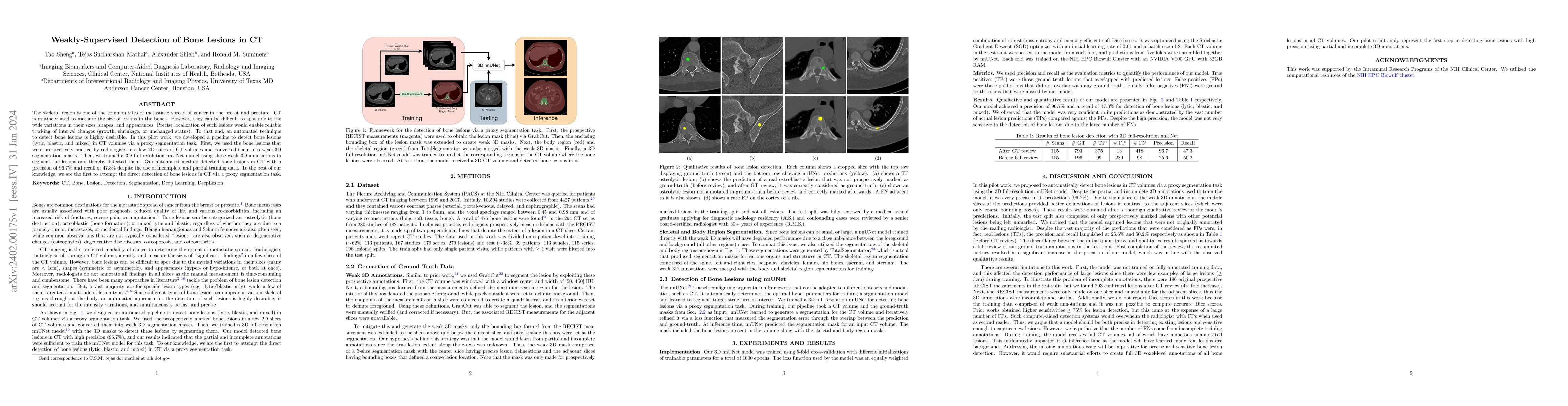
The skeletal region is one of the common sites of metastatic spread of cancer in the breast and prostate. CT is routinely used to measure the size of lesions in the bones. However, they can be diffi...
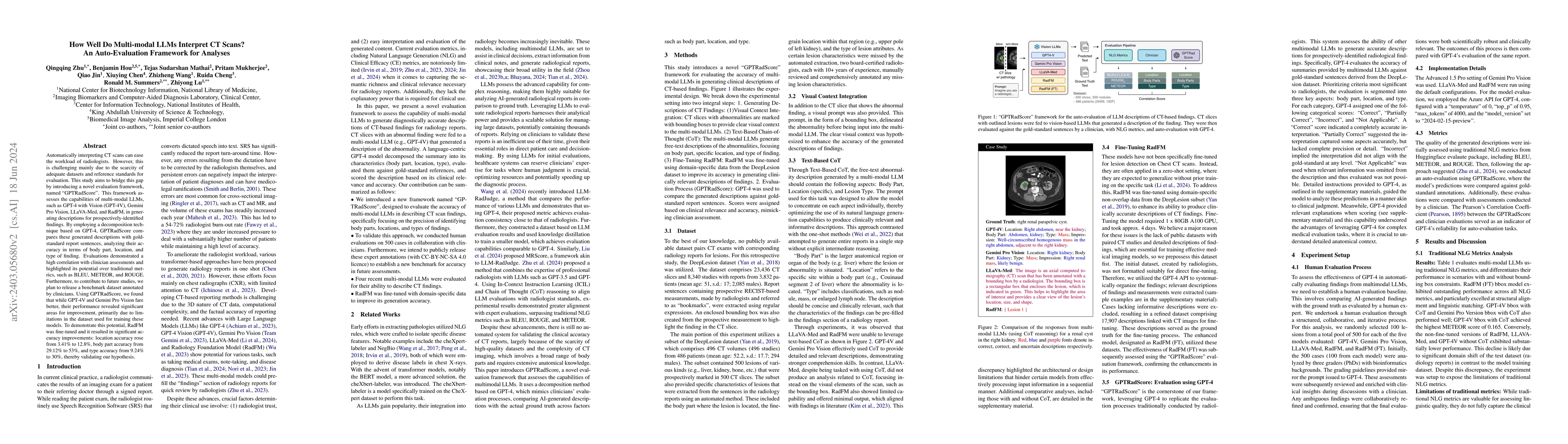
Recent studies indicate that Generative Pre-trained Transformer 4 with Vision (GPT-4V) outperforms human physicians in medical challenge tasks. However, these evaluations primarily focused on the ac...
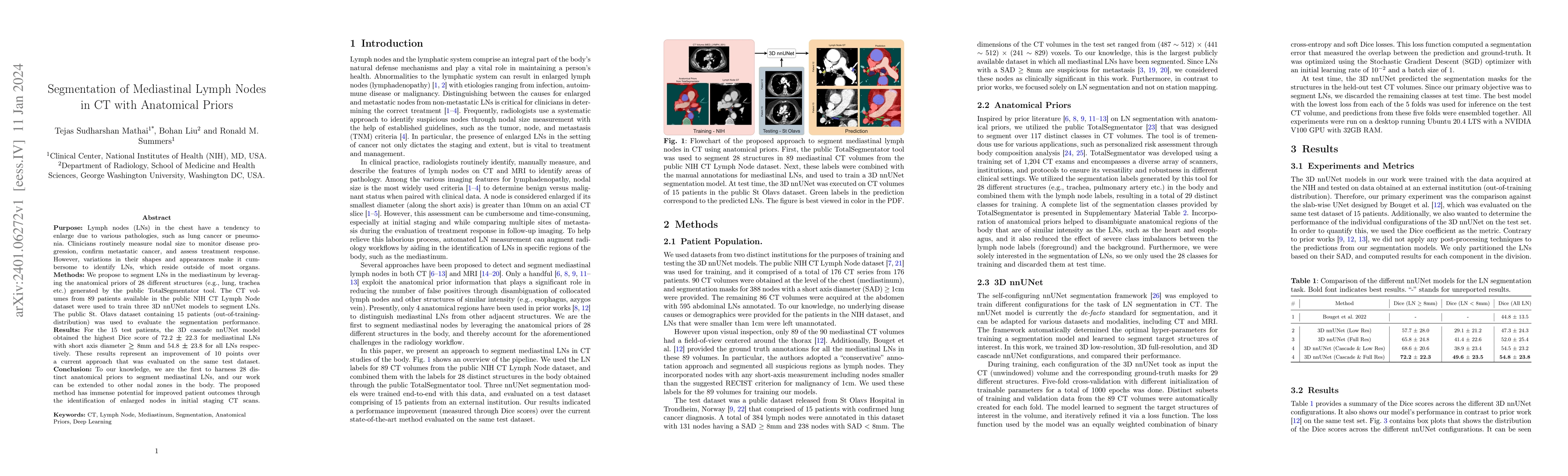
Purpose: Lymph nodes (LNs) in the chest have a tendency to enlarge due to various pathologies, such as lung cancer or pneumonia. Clinicians routinely measure nodal size to monitor disease progressio...
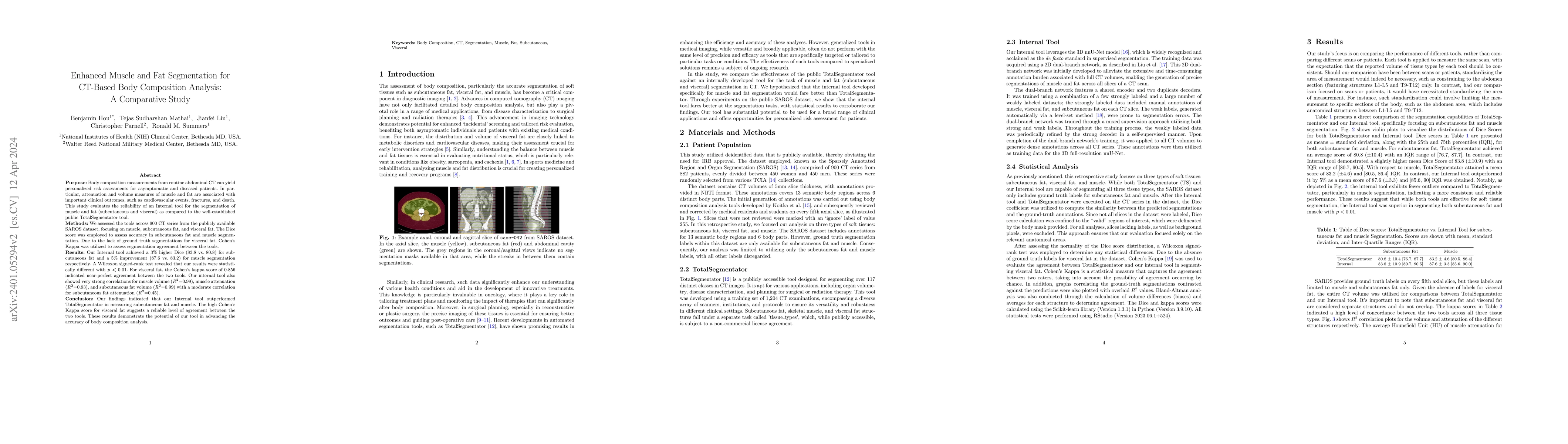
Purpose: Body composition measurements from routine abdominal CT can yield personalized risk assessments for asymptomatic and diseased patients. In particular, attenuation and volume measures of mus...
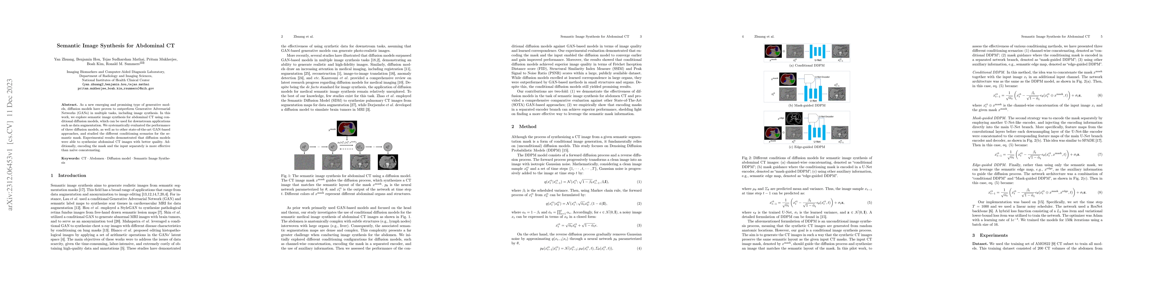
As a new emerging and promising type of generative models, diffusion models have proven to outperform Generative Adversarial Networks (GANs) in multiple tasks, including image synthesis. In this wor...
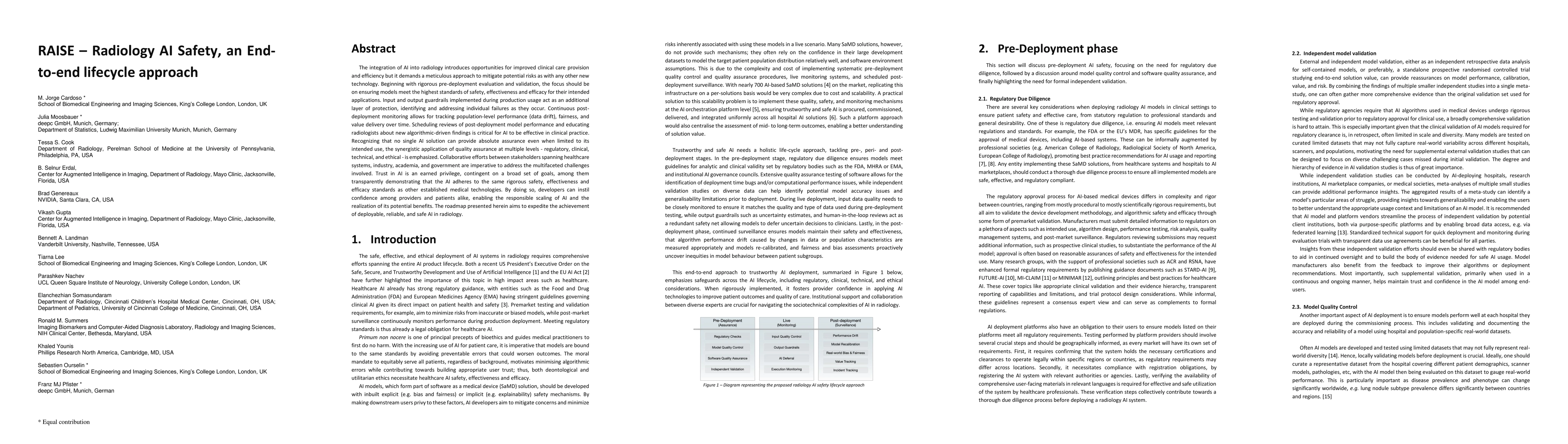
The integration of AI into radiology introduces opportunities for improved clinical care provision and efficiency but it demands a meticulous approach to mitigate potential risks as with any other n...
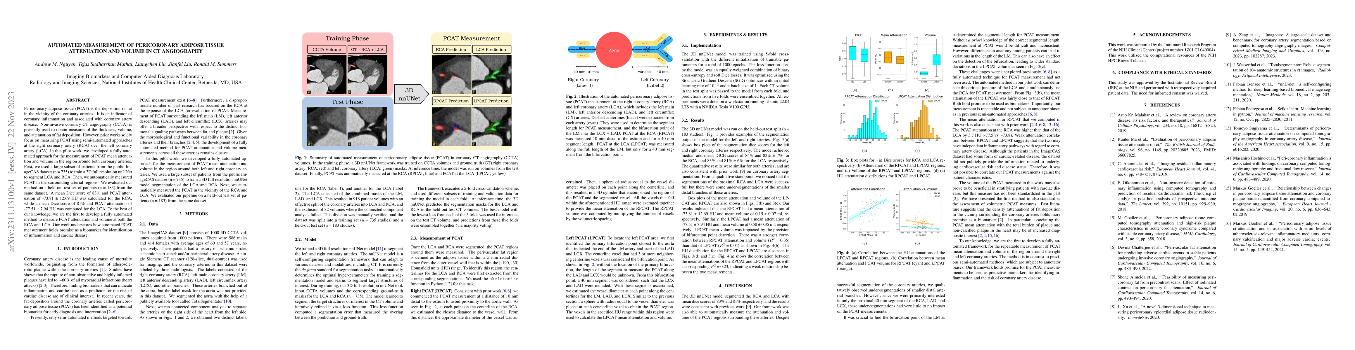
Pericoronary adipose tissue (PCAT) is the deposition of fat in the vicinity of the coronary arteries. It is an indicator of coronary inflammation and associated with coronary artery disease. Non-inv...
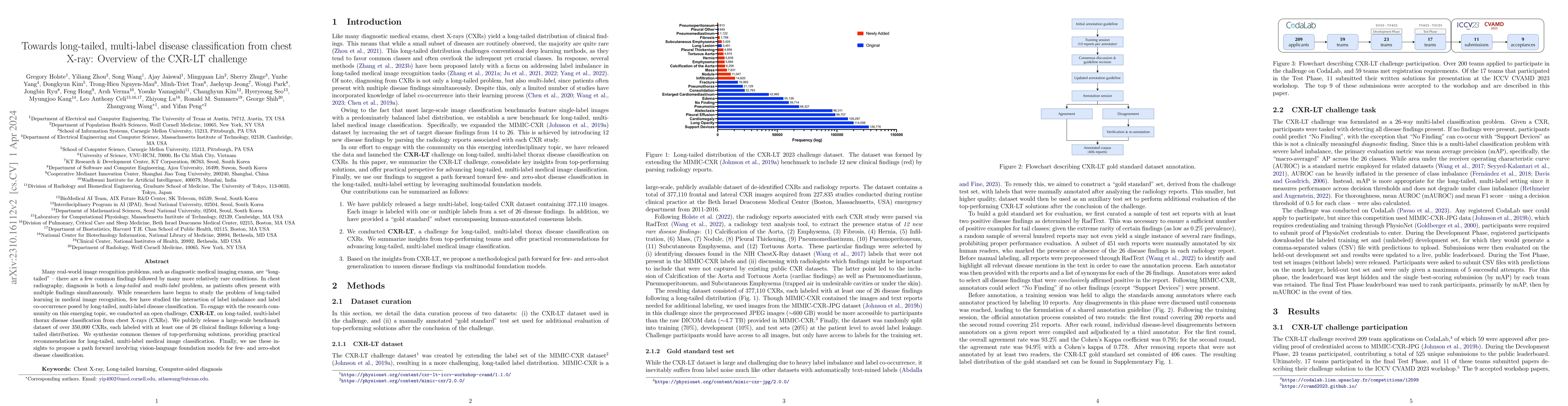
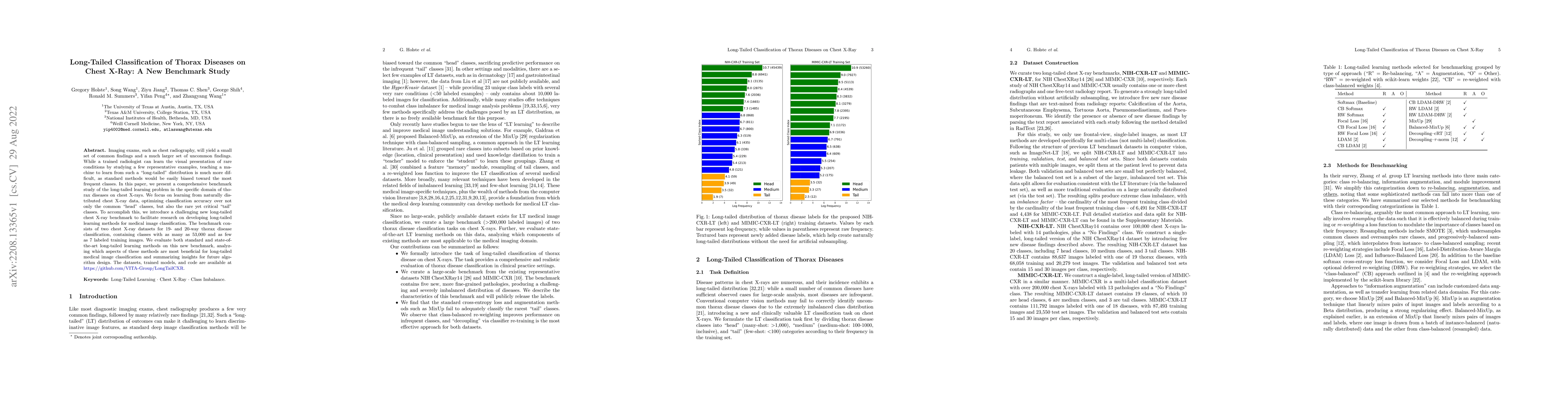
Many real-world image recognition problems, such as diagnostic medical imaging exams, are "long-tailed" $\unicode{x2013}$ there are a few common findings followed by many more relatively rare condit...

Patients undergoing chest X-rays (CXR) often endure multiple lung diseases. When evaluating a patient's condition, due to the complex pathologies, subtle texture changes of different lung lesions in...
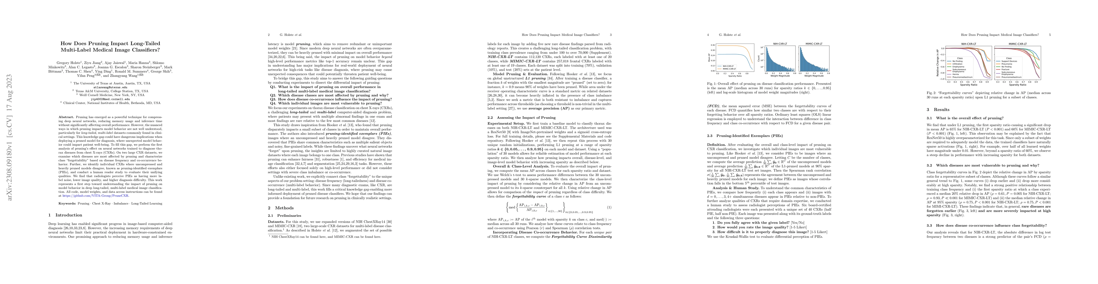
Pruning has emerged as a powerful technique for compressing deep neural networks, reducing memory usage and inference time without significantly affecting overall performance. However, the nuanced w...
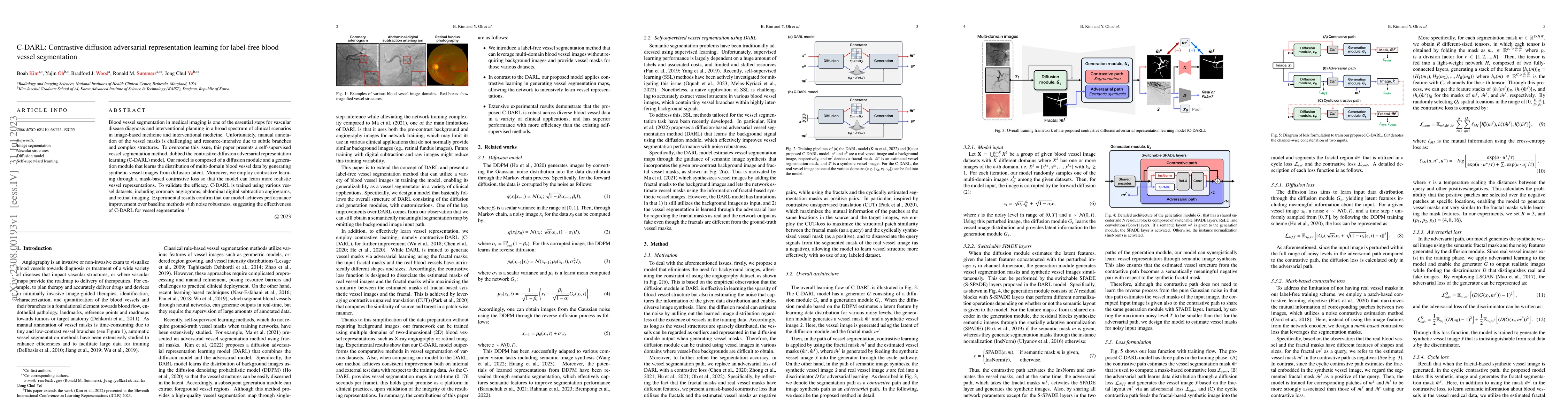
Blood vessel segmentation in medical imaging is one of the essential steps for vascular disease diagnosis and interventional planning in a broad spectrum of clinical scenarios in image-based medicin...
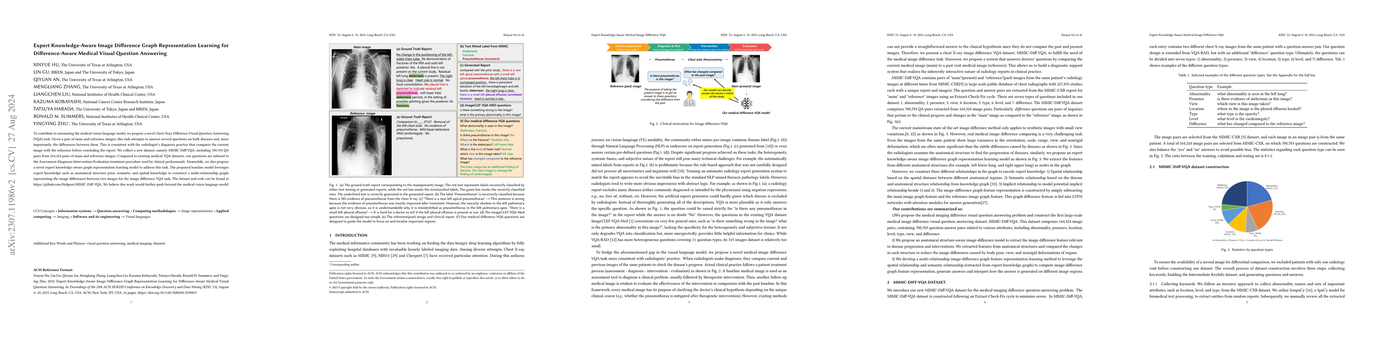
To contribute to automating the medical vision-language model, we propose a novel Chest-Xray Difference Visual Question Answering (VQA) task. Given a pair of main and reference images, this task att...
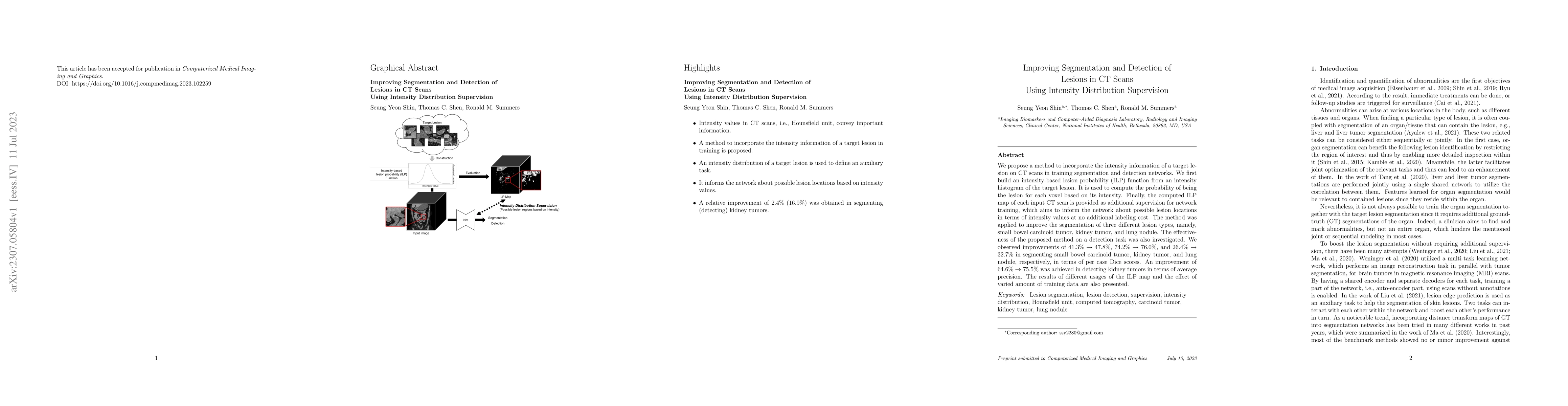
We propose a method to incorporate the intensity information of a target lesion on CT scans in training segmentation and detection networks. We first build an intensity-based lesion probability (ILP...
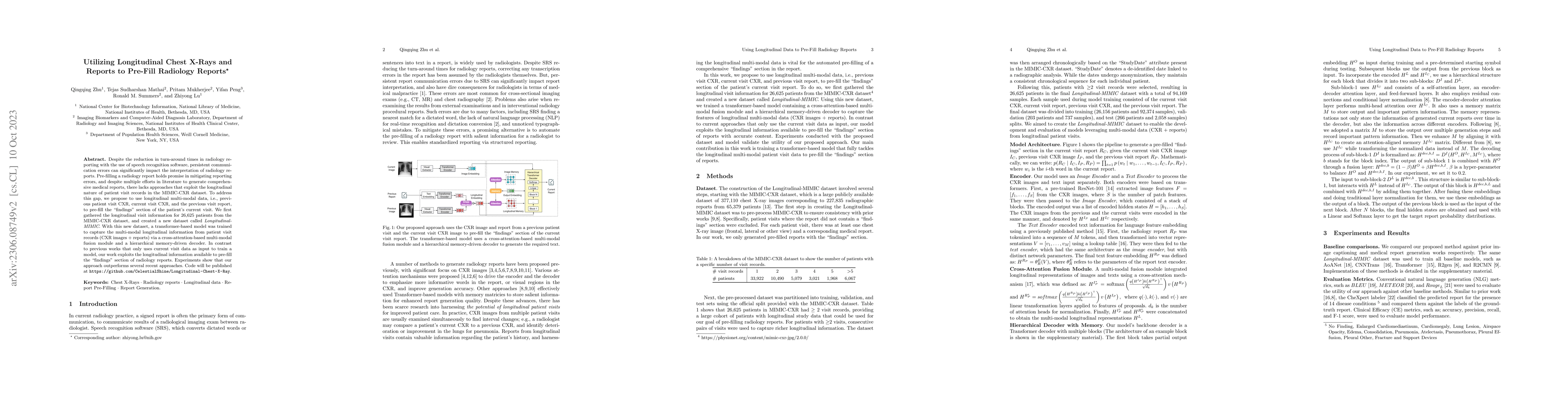
Despite the reduction in turn-around times in radiology reports with the use of speech recognition software, persistent communication errors can significantly impact the interpretation of the radiol...
Validation metrics are key for the reliable tracking of scientific progress and for bridging the current chasm between artificial intelligence (AI) research and its translation into practice. Howeve...
Imaging exams, such as chest radiography, will yield a small set of common findings and a much larger set of uncommon findings. While a trained radiologist can learn the visual presentation of rare ...
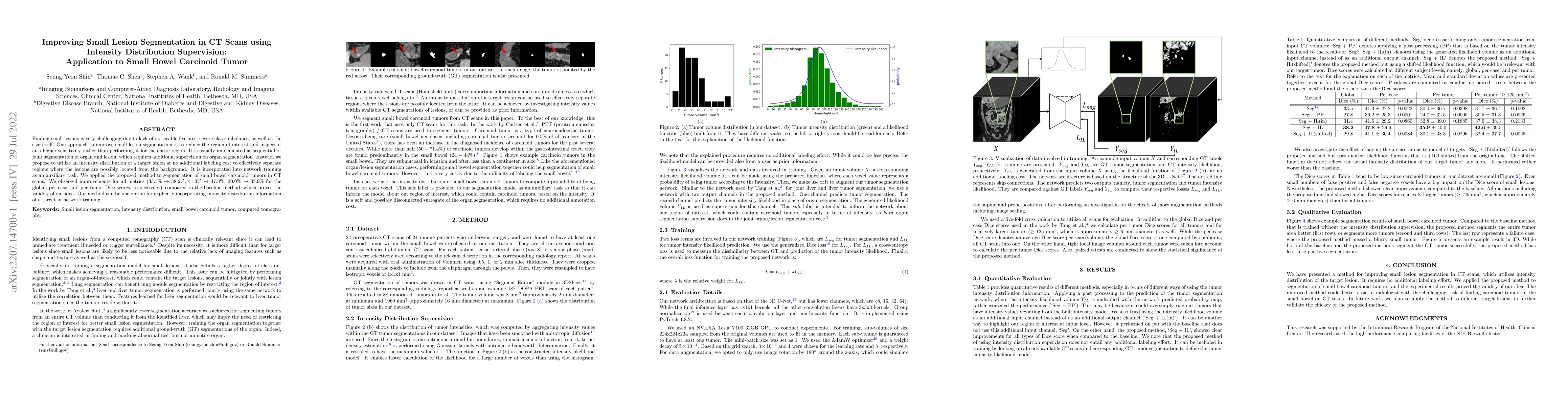
Finding small lesions is very challenging due to lack of noticeable features, severe class imbalance, as well as the size itself. One approach to improve small lesion segmentation is to reduce the r...
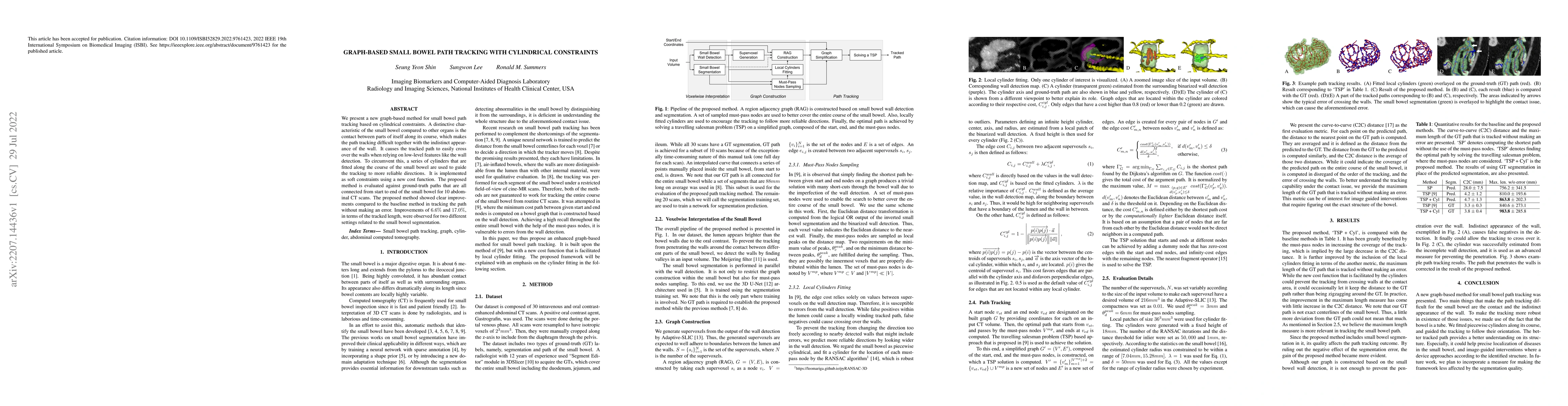
We present a new graph-based method for small bowel path tracking based on cylindrical constraints. A distinctive characteristic of the small bowel compared to other organs is the contact between pa...
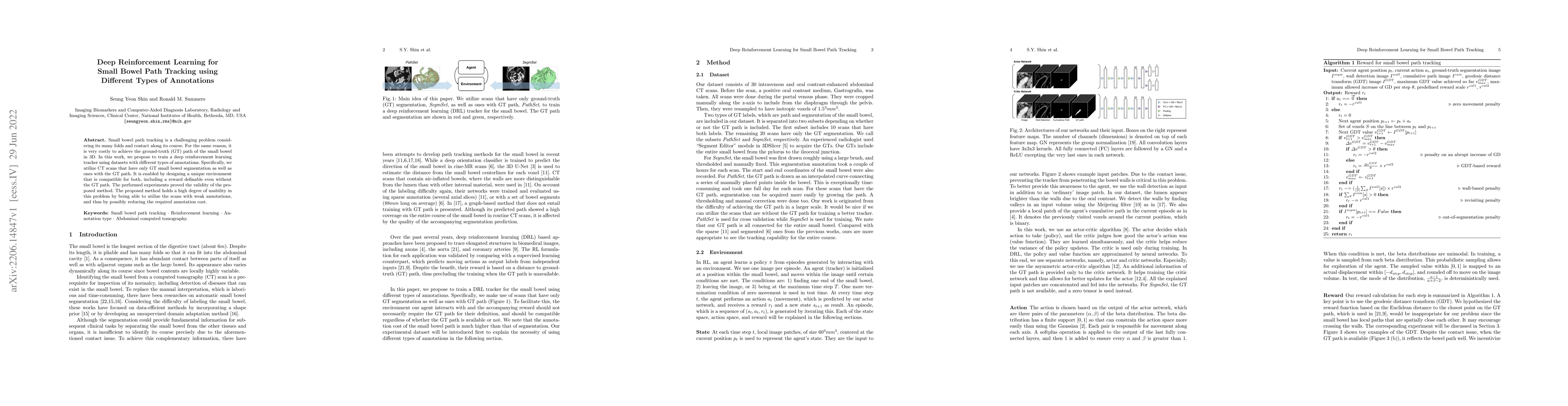
Small bowel path tracking is a challenging problem considering its many folds and contact along its course. For the same reason, it is very costly to achieve the ground-truth (GT) path of the small ...
Increasing evidence shows that flaws in machine learning (ML) algorithm validation are an underestimated global problem. Particularly in automatic biomedical image analysis, chosen performance metri...
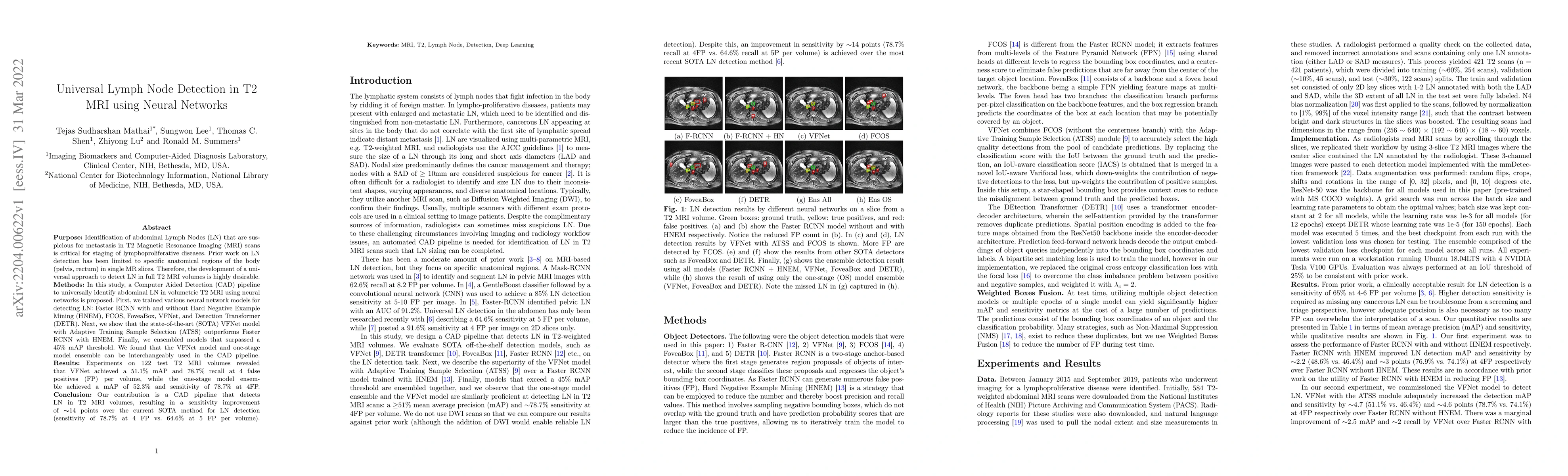
Purpose: Identification of abdominal Lymph Nodes (LN) that are suspicious for metastasis in T2 Magnetic Resonance Imaging (MRI) scans is critical for staging of lymphoproliferative diseases. Prior w...
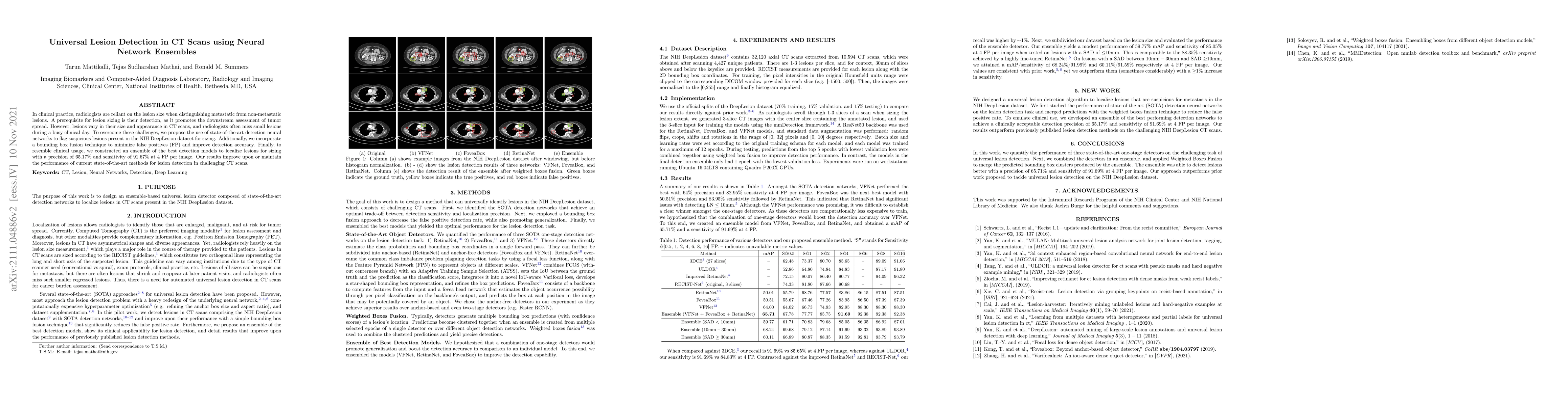
In clinical practice, radiologists are reliant on the lesion size when distinguishing metastatic from non-metastatic lesions. A prerequisite for lesion sizing is their detection, as it promotes the ...
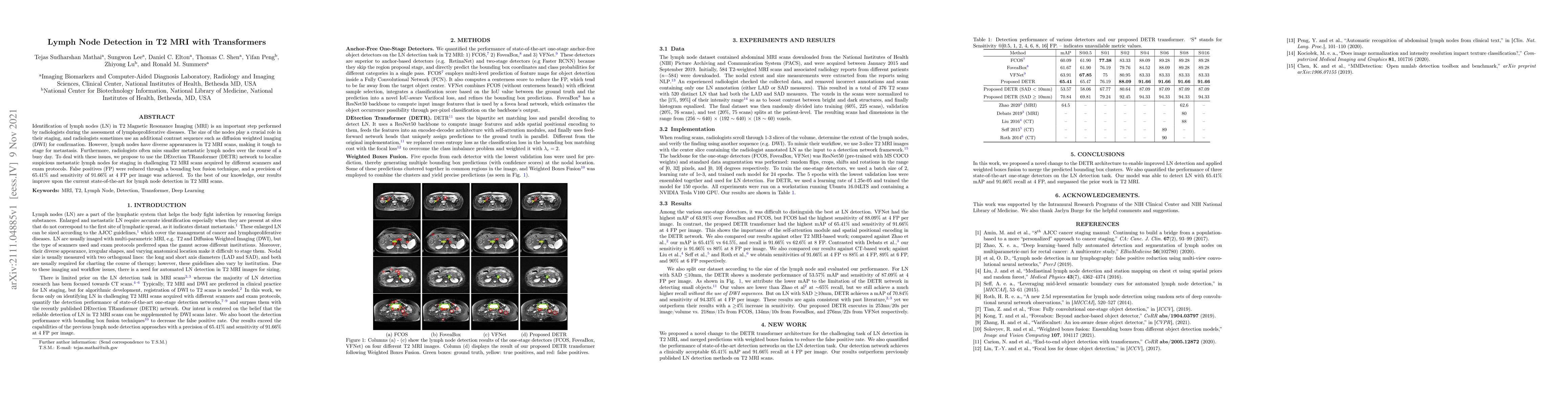
Identification of lymph nodes (LN) in T2 Magnetic Resonance Imaging (MRI) is an important step performed by radiologists during the assessment of lymphoproliferative diseases. The size of the nodes ...
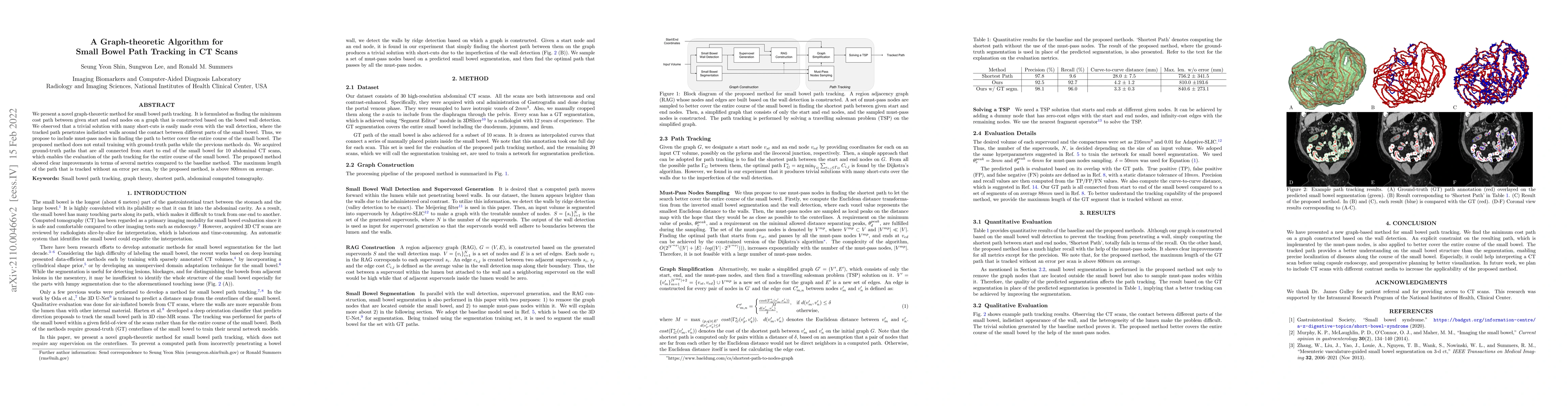
We present a novel graph-theoretic method for small bowel path tracking. It is formulated as finding the minimum cost path between given start and end nodes on a graph that is constructed based on t...
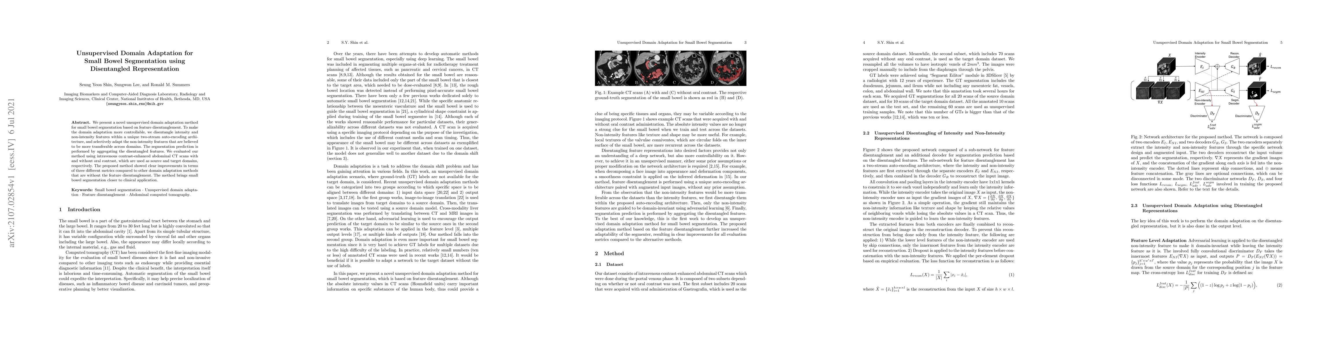
We present a novel unsupervised domain adaptation method for small bowel segmentation based on feature disentanglement. To make the domain adaptation more controllable, we disentangle intensity and ...
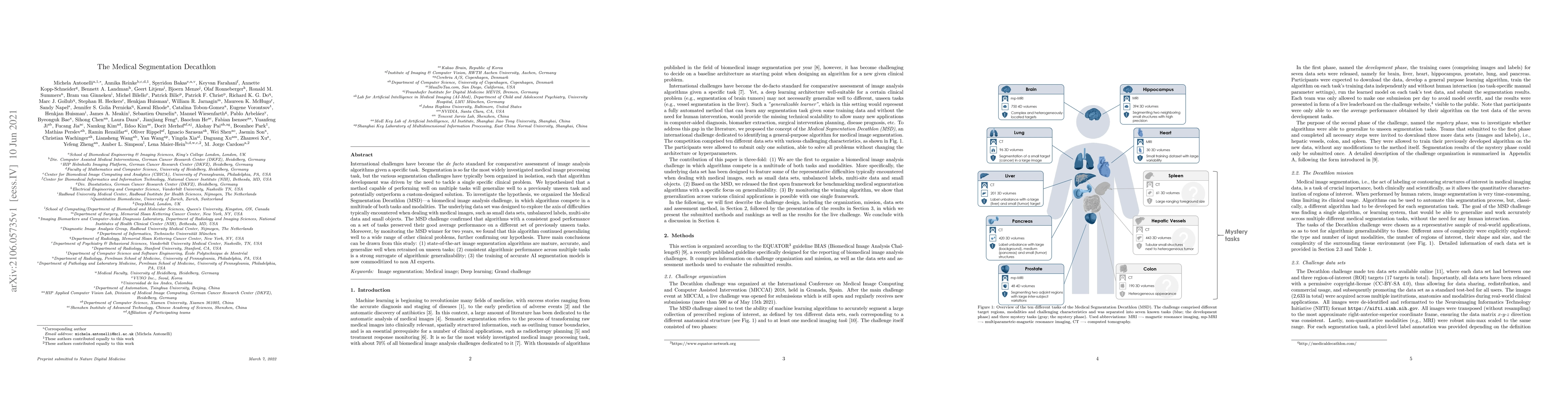
International challenges have become the de facto standard for comparative assessment of image analysis algorithms given a specific task. Segmentation is so far the most widely investigated medical ...
While the importance of automatic image analysis is continuously increasing, recent meta-research revealed major flaws with respect to algorithm validation. Performance metrics are particularly key ...
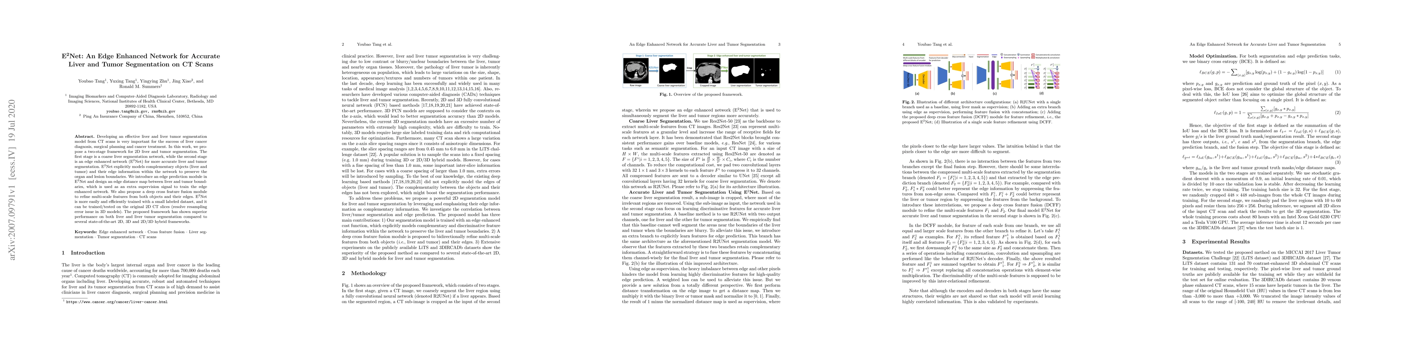
Developing an effective liver and liver tumor segmentation model from CT scans is very important for the success of liver cancer diagnosis, surgical planning and cancer treatment. In this work, we p...
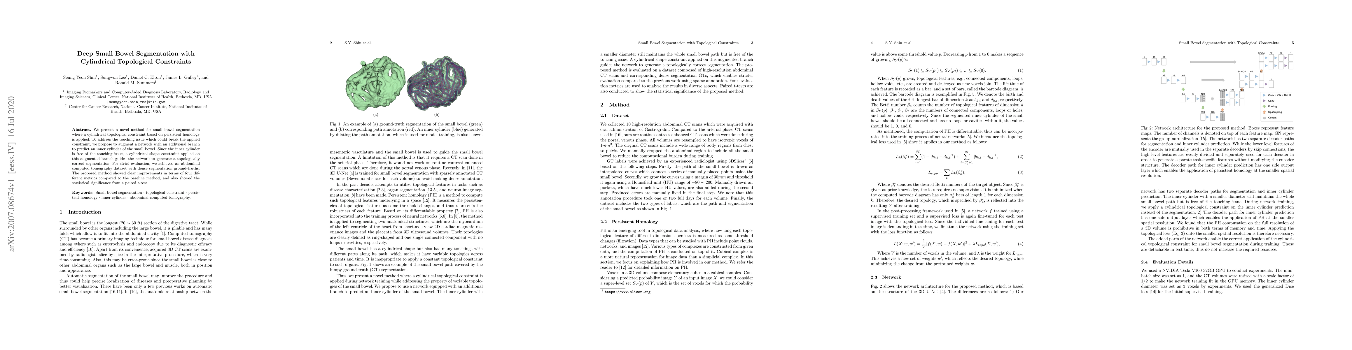
We present a novel method for small bowel segmentation where a cylindrical topological constraint based on persistent homology is applied. To address the touching issue which could break the applied...
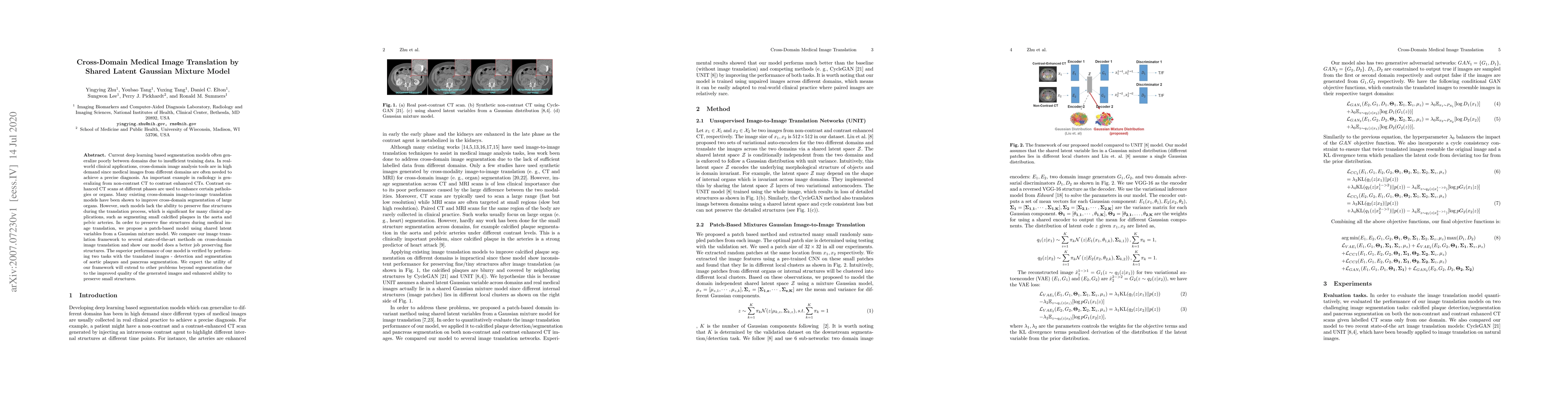
Current deep learning based segmentation models often generalize poorly between domains due to insufficient training data. In real-world clinical applications, cross-domain image analysis tools are ...
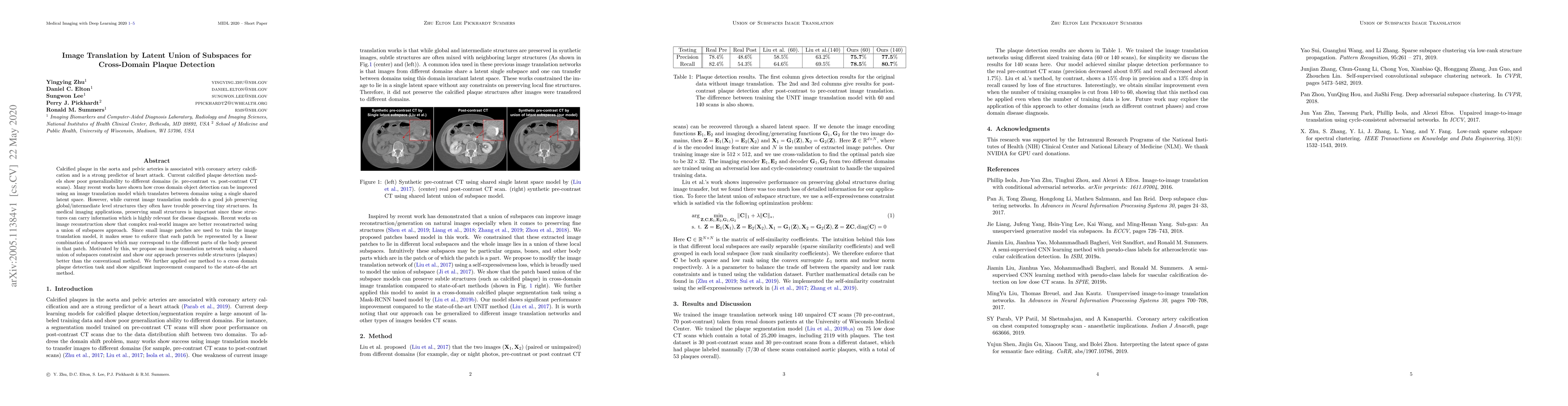
Calcified plaque in the aorta and pelvic arteries is associated with coronary artery calcification and is a strong predictor of heart attack. Current calcified plaque detection models show poor gene...
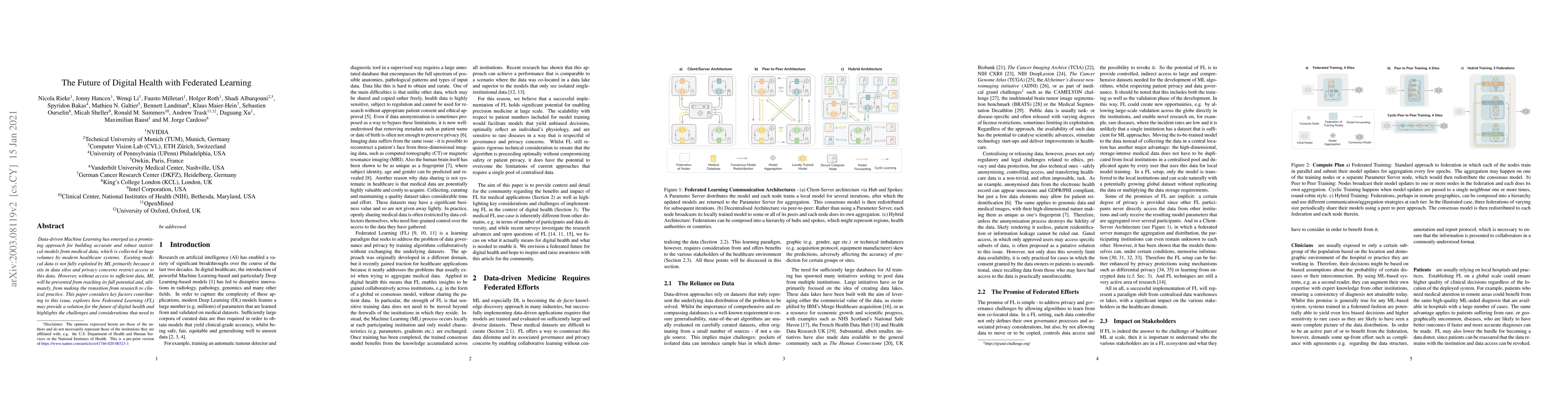
Data-driven Machine Learning has emerged as a promising approach for building accurate and robust statistical models from medical data, which is collected in huge volumes by modern healthcare system...
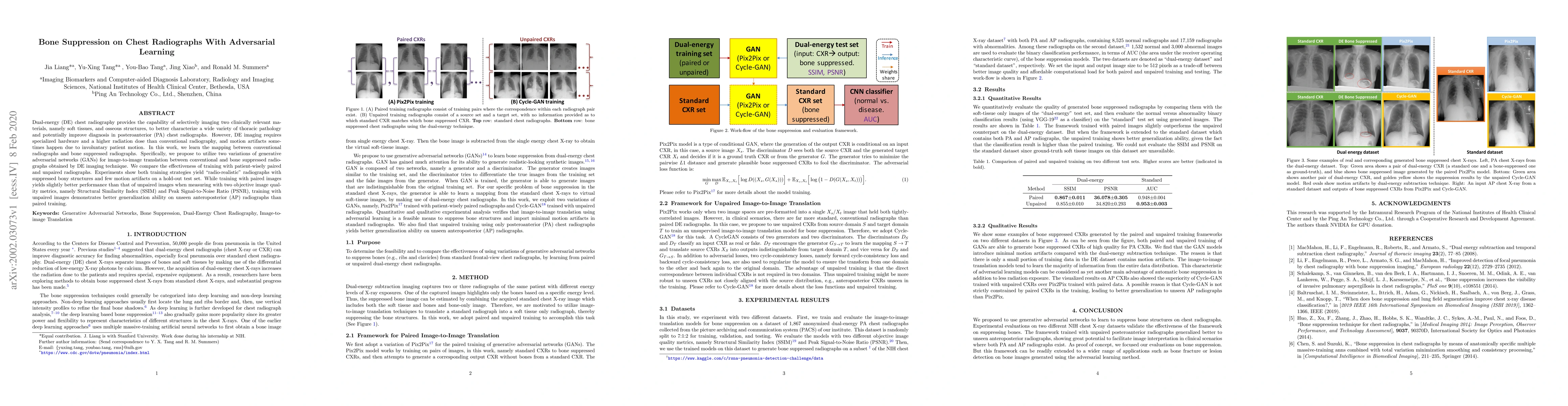
Dual-energy (DE) chest radiography provides the capability of selectively imaging two clinically relevant materials, namely soft tissues, and osseous structures, to better characterize a wide variet...
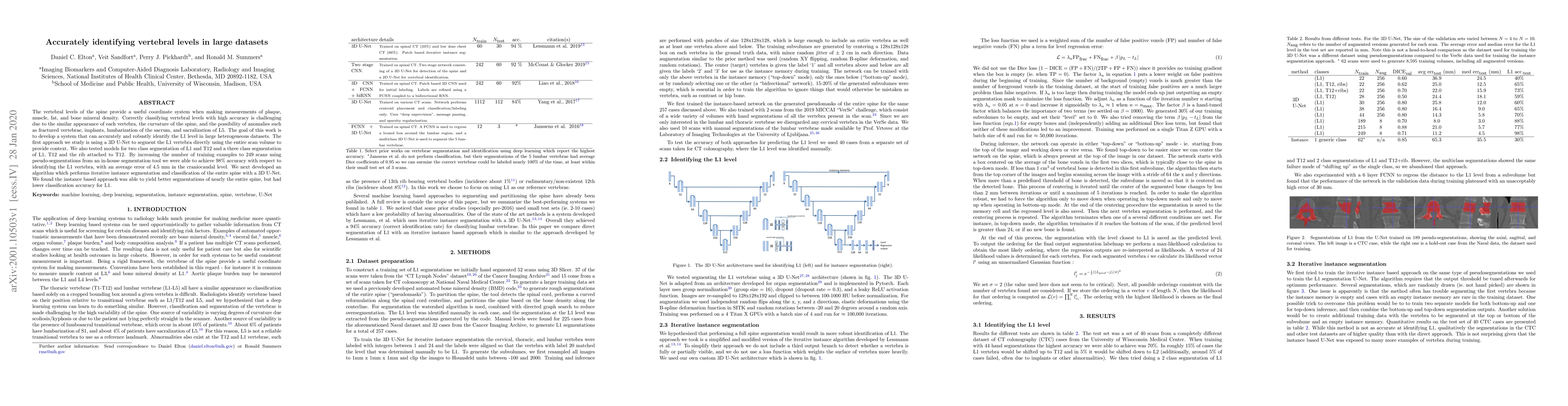
The vertebral levels of the spine provide a useful coordinate system when making measurements of plaque, muscle, fat, and bone mineral density. Correctly classifying vertebral levels with high accur...
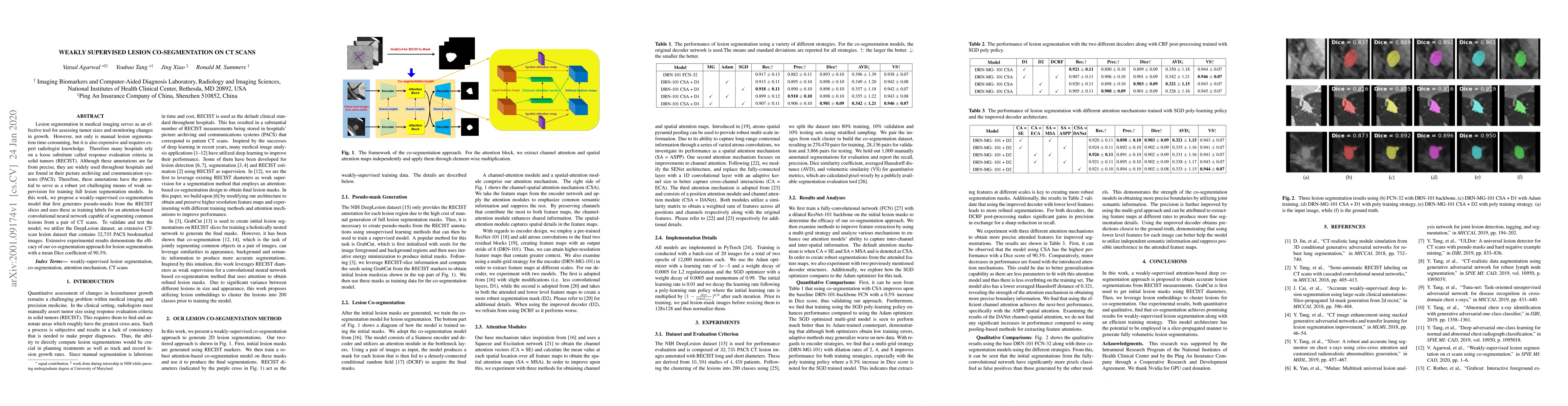
Lesion segmentation in medical imaging serves as an effective tool for assessing tumor sizes and monitoring changes in growth. However, not only is manual lesion segmentation time-consuming, but it ...
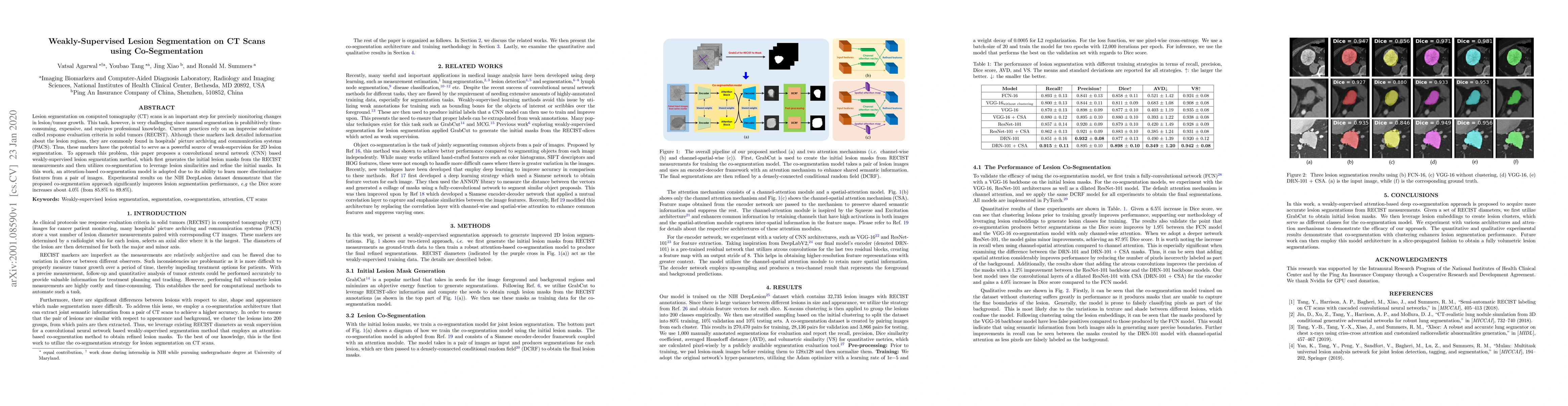
Lesion segmentation on computed tomography (CT) scans is an important step for precisely monitoring changes in lesion/tumor growth. This task, however, is very challenging since manual segmentation ...
Purpose: To systematically investigate the influence of various data consistency layers, (semi-)supervised learning and ensembling strategies, defined in a $\Sigma$-net, for accelerated parallel MR ...
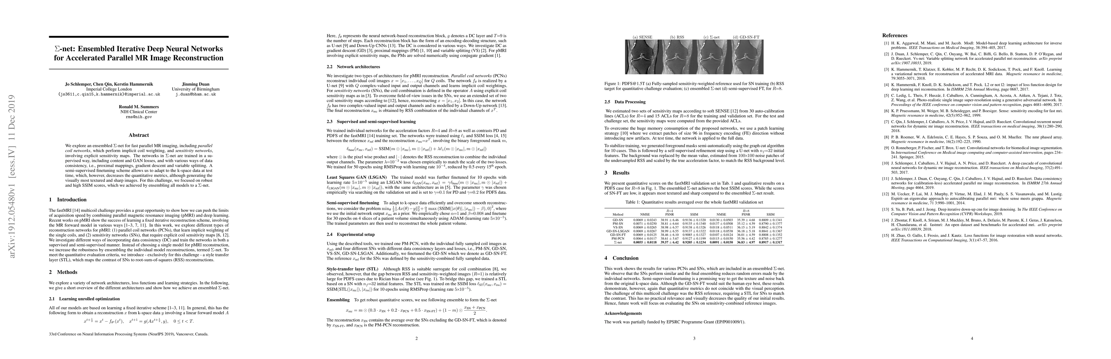
We explore an ensembled $\Sigma$-net for fast parallel MR imaging, including parallel coil networks, which perform implicit coil weighting, and sensitivity networks, involving explicit sensitivity m...
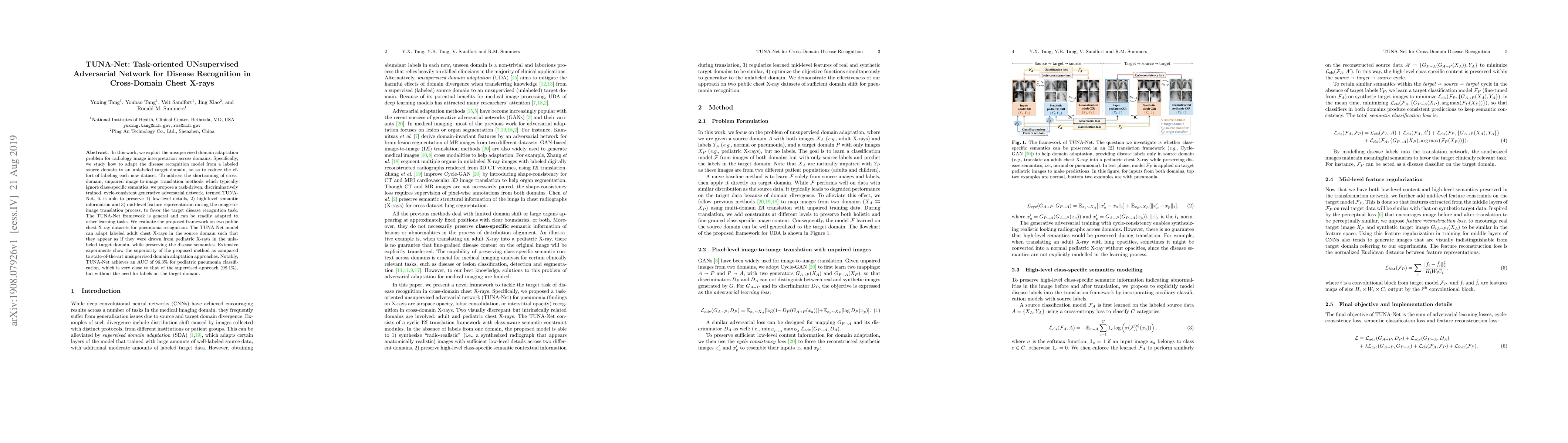
In this work, we exploit the unsupervised domain adaptation problem for radiology image interpretation across domains. Specifically, we study how to adapt the disease recognition model from a labele...
When reading medical images such as a computed tomography (CT) scan, radiologists generally search across the image to find lesions, characterize and measure them, and then describe them in the radi...
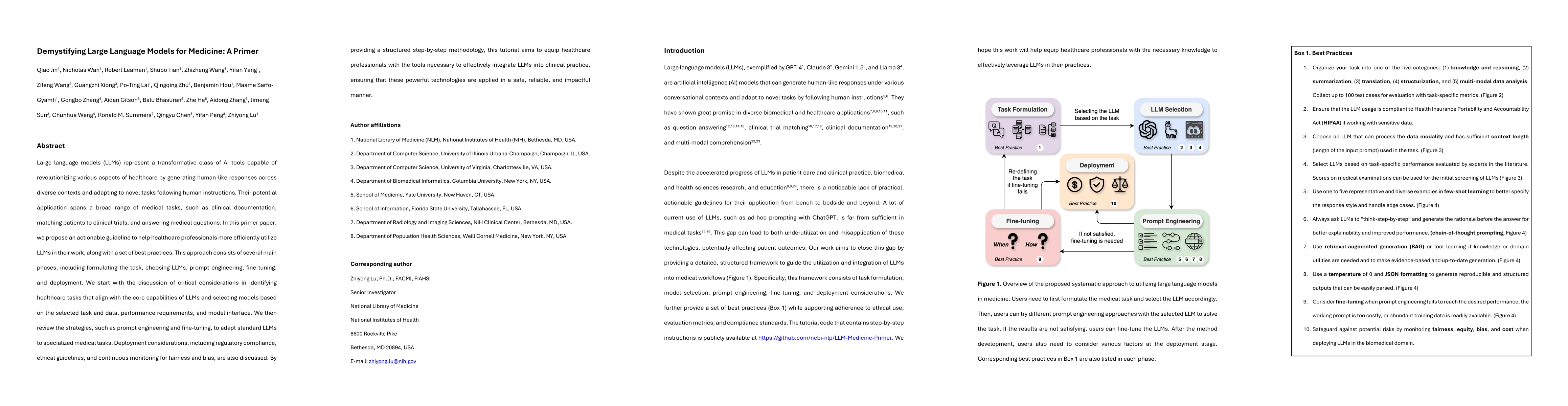
Large language models (LLMs) represent a transformative class of AI tools capable of revolutionizing various aspects of healthcare by generating human-like responses across diverse contexts and adapti...
Purpose: The purpose of this study is to harness the efficiency of a 2D foundation model to develop a robust phase classifier that is resilient to domain shifts. Materials and Methods: This retrospe...
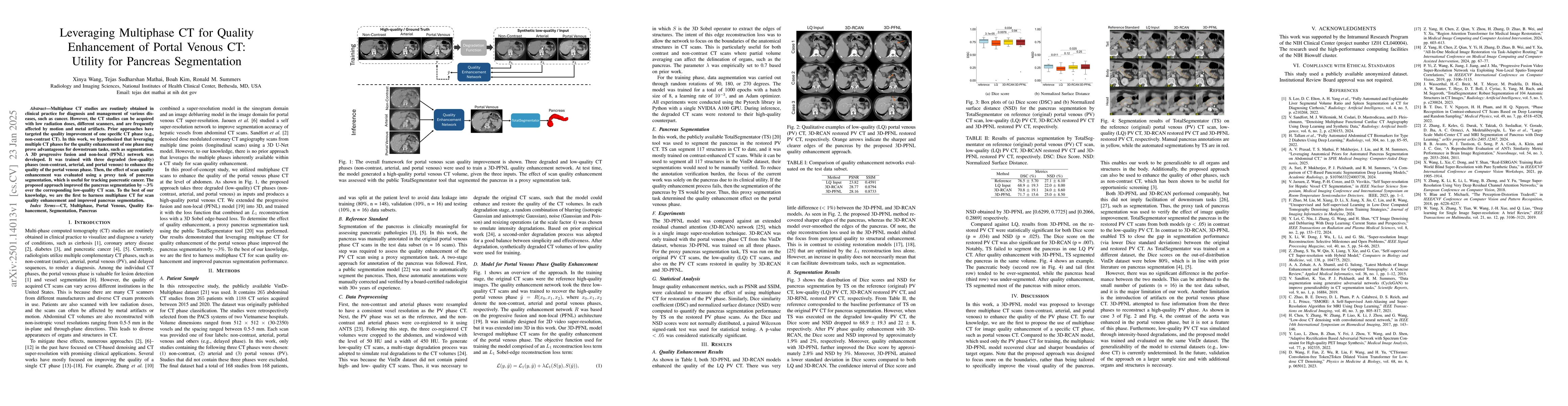
Multiphase CT studies are routinely obtained in clinical practice for diagnosis and management of various diseases, such as cancer. However, the CT studies can be acquired with low radiation doses, di...
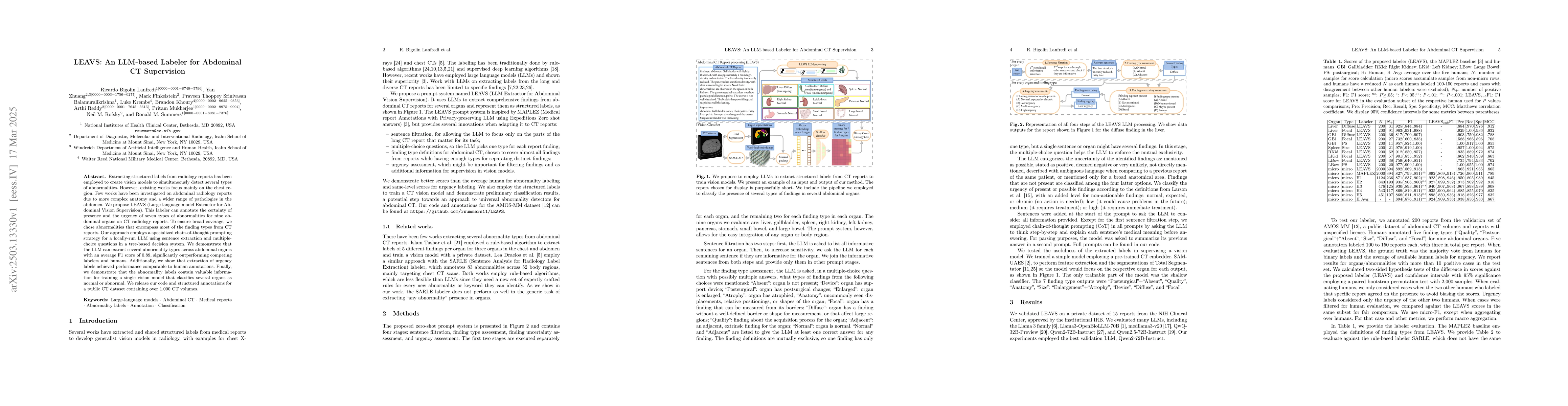
Extracting structured labels from radiology reports has been employed to create vision models to simultaneously detect several types of abnormalities. However, existing works focus mainly on the chest...
Precision medicine in the quantitative management of chronic diseases and oncology would be greatly improved if the Computed Tomography (CT) scan of any patient could be segmented, parsed and analyzed...
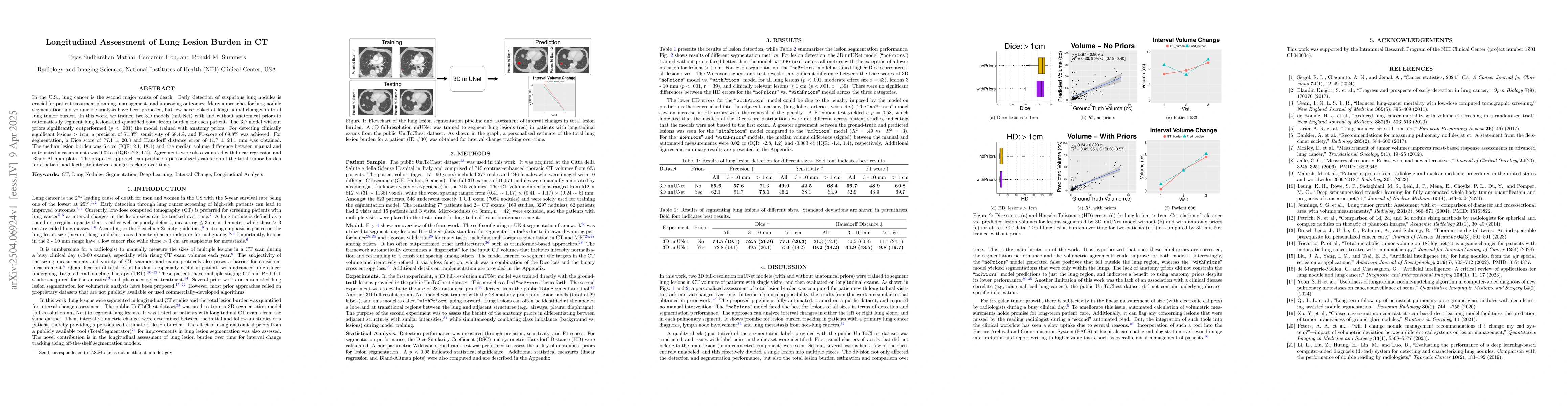
In the U.S., lung cancer is the second major cause of death. Early detection of suspicious lung nodules is crucial for patient treatment planning, management, and improving outcomes. Many approaches f...
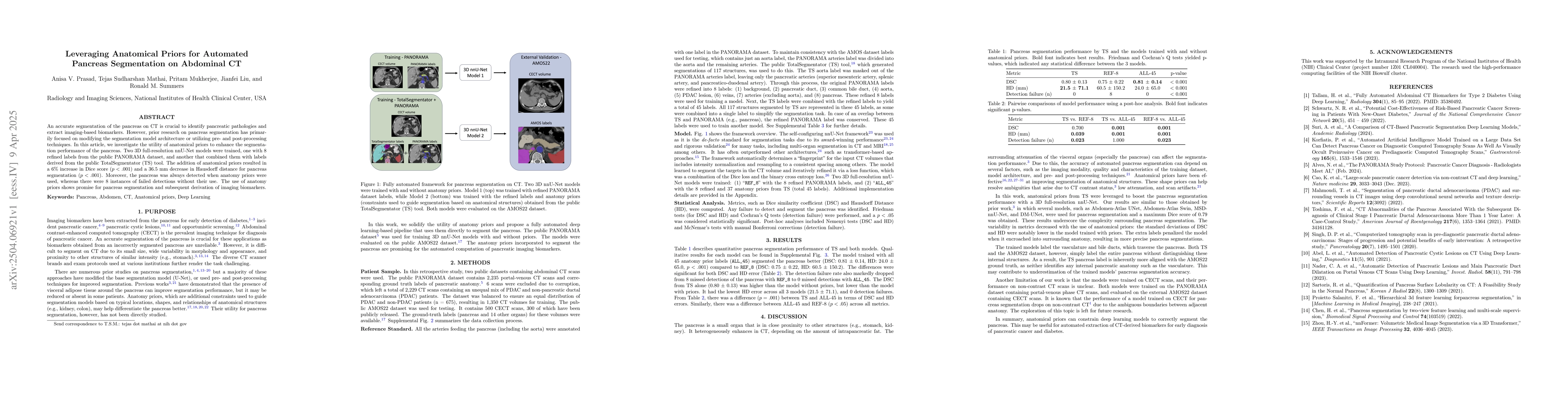
An accurate segmentation of the pancreas on CT is crucial to identify pancreatic pathologies and extract imaging-based biomarkers. However, prior research on pancreas segmentation has primarily focuse...
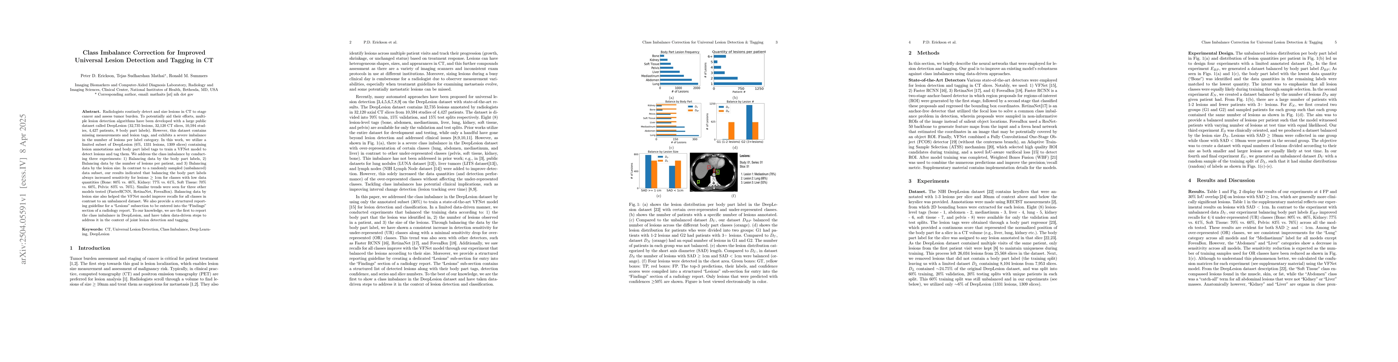
Radiologists routinely detect and size lesions in CT to stage cancer and assess tumor burden. To potentially aid their efforts, multiple lesion detection algorithms have been developed with a large pu...
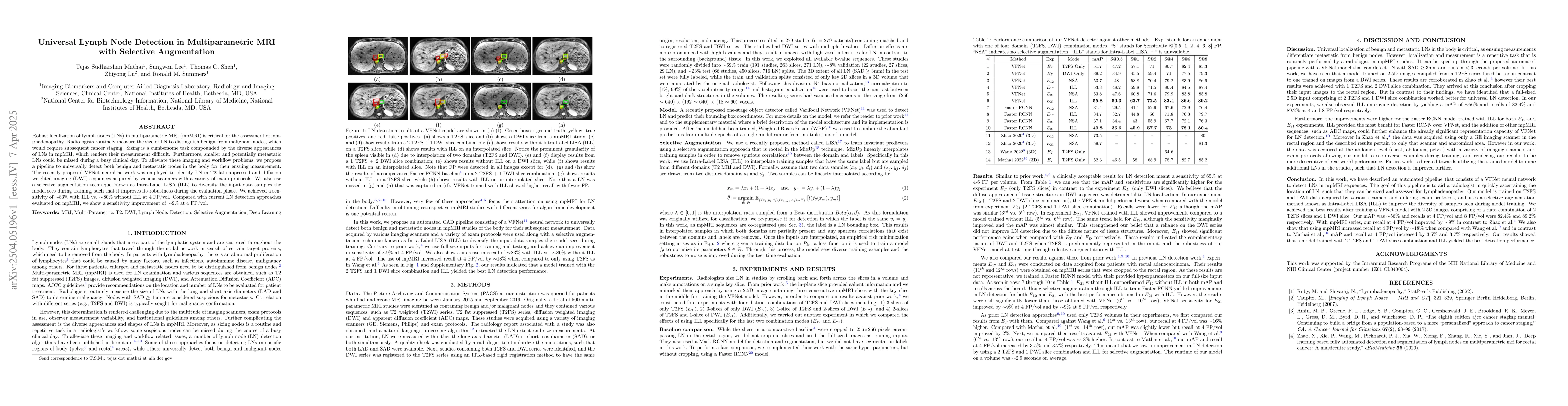
Robust localization of lymph nodes (LNs) in multiparametric MRI (mpMRI) is critical for the assessment of lymphadenopathy. Radiologists routinely measure the size of LN to distinguish benign from mali...
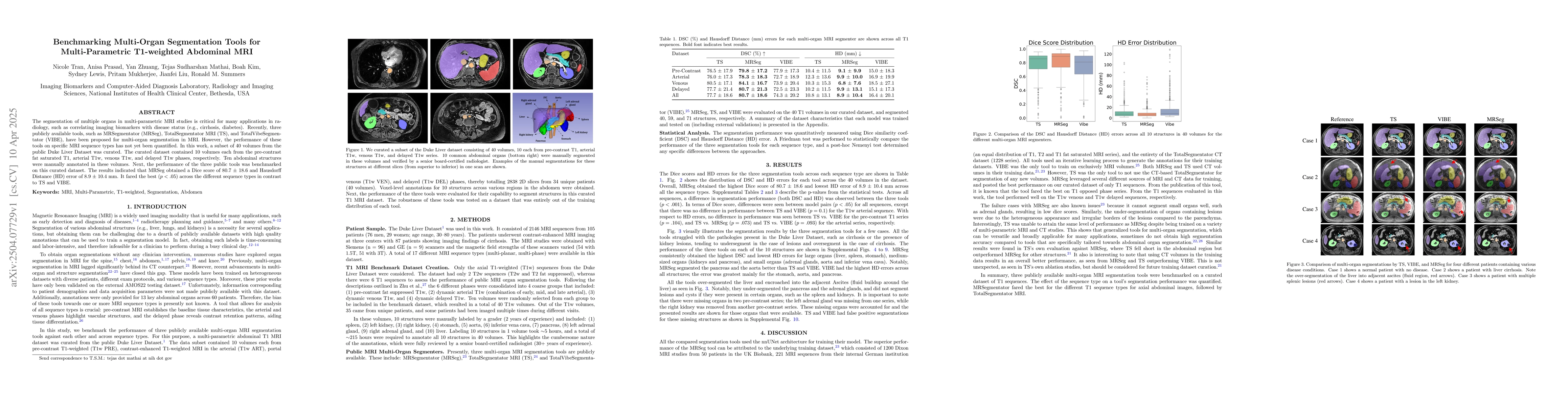
The segmentation of multiple organs in multi-parametric MRI studies is critical for many applications in radiology, such as correlating imaging biomarkers with disease status (e.g., cirrhosis, diabete...
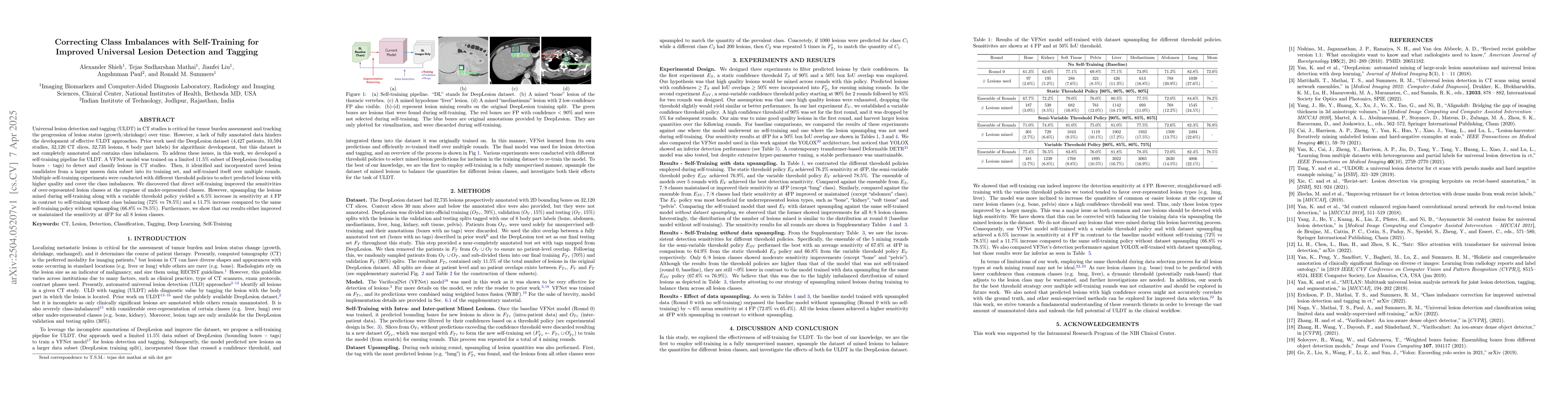
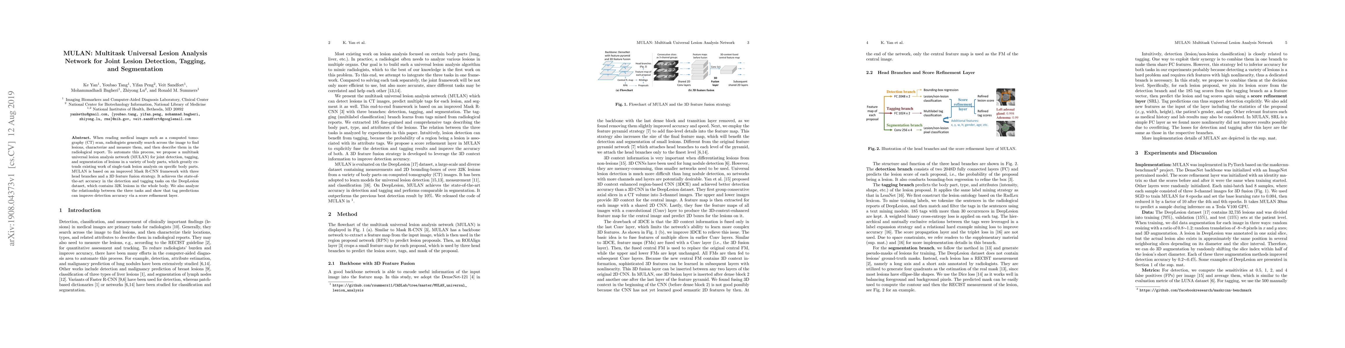
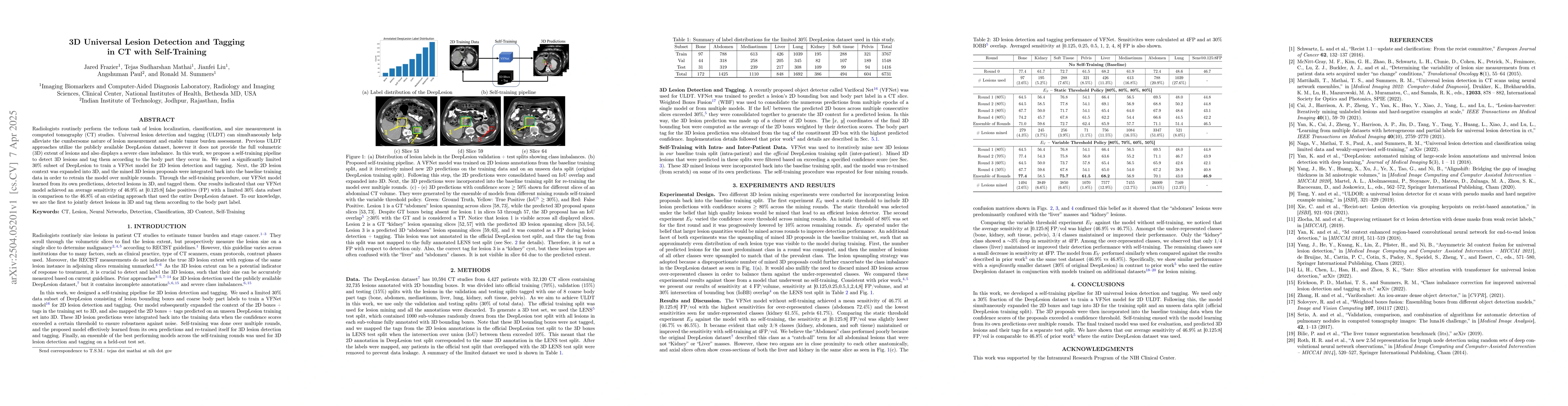
Universal lesion detection and tagging (ULDT) in CT studies is critical for tumor burden assessment and tracking the progression of lesion status (growth/shrinkage) over time. However, a lack of fully...
Radiologists routinely perform the tedious task of lesion localization, classification, and size measurement in computed tomography (CT) studies. Universal lesion detection and tagging (ULDT) can simu...

The CXR-LT series is a community-driven initiative designed to enhance lung disease classification using chest X-rays (CXR). It tackles challenges in open long-tailed lung disease classification and e...
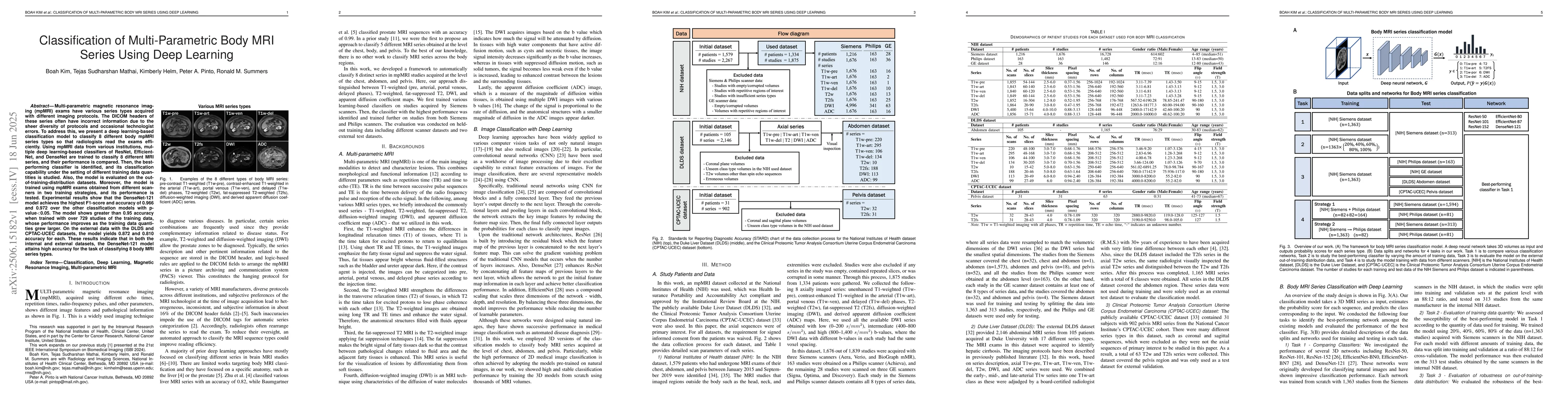
Multi-parametric magnetic resonance imaging (mpMRI) exams have various series types acquired with different imaging protocols. The DICOM headers of these series often have incorrect information due to...
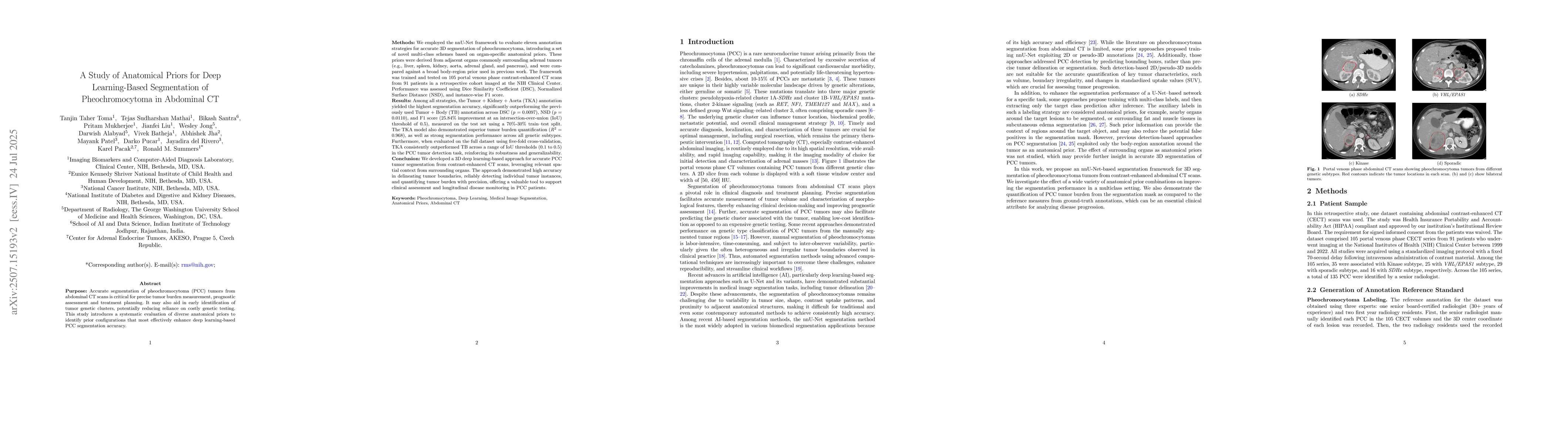
Accurate segmentation of pheochromocytoma (PCC) in abdominal CT scans is essential for tumor burden estimation, prognosis, and treatment planning. It may also help infer genetic clusters, reducing rel...
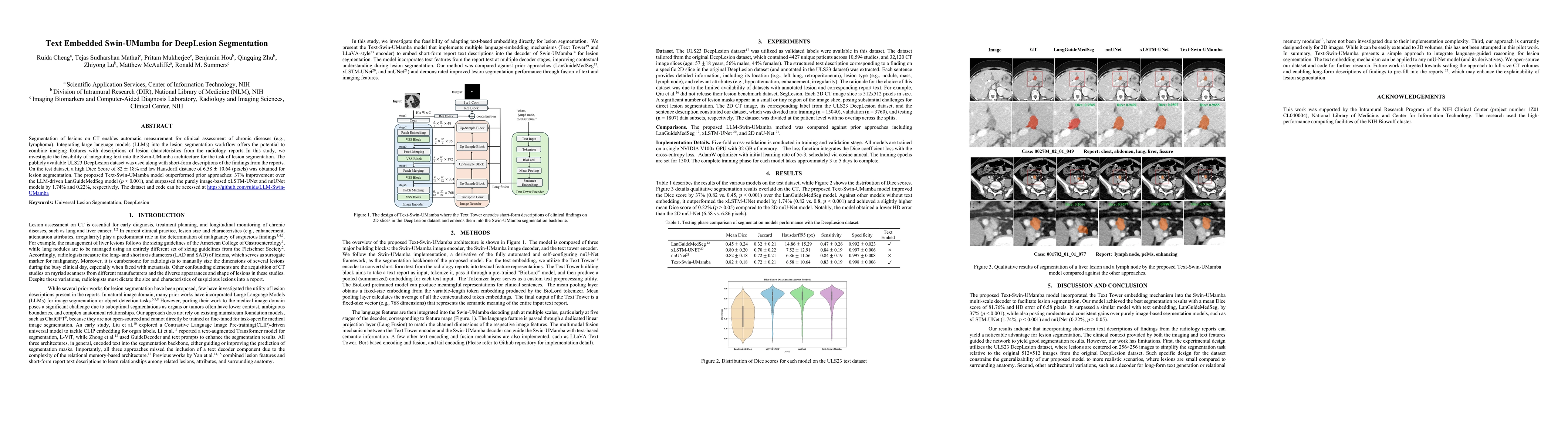
Segmentation of lesions on CT enables automatic measurement for clinical assessment of chronic diseases (e.g., lymphoma). Integrating large language models (LLMs) into the lesion segmentation workflow...
Type 2 Diabetes Mellitus (T2DM) is a chronic metabolic disease that affects millions of people worldwide. Early detection is crucial as it can alter pancreas function through morphological changes and...
Artificial intelligence (AI) can automatically delineate lesions on computed tomography (CT) and generate radiology report content, yet progress is limited by the scarcity of publicly available CT dat...
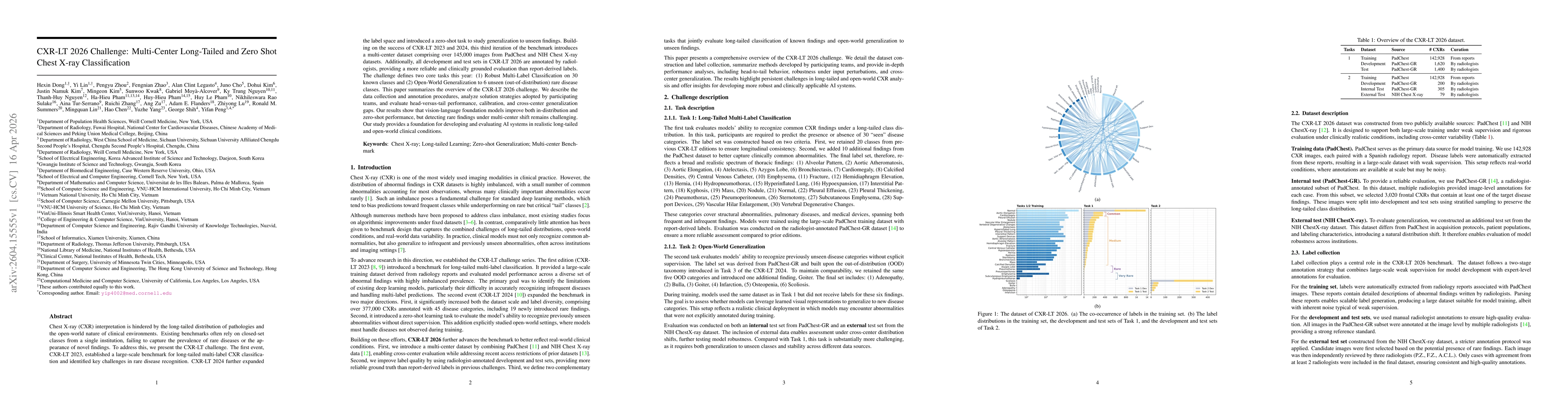
Chest X-ray (CXR) interpretation is hindered by the long-tailed distribution of pathologies and the open-world nature of clinical environments. Existing benchmarks often rely on closed-set classes fro...