Universal Lymph Node Detection in T2 MRI using Neural Networks
Publication
Metrics
AI Quick Summary
This paper proposes a Computer Aided Detection (CAD) pipeline using neural networks to universally identify abdominal lymph nodes in full T2 MRI volumes. The VFNet model with Adaptive Training Sample Selection achieved the best results, showing a 51.1% mean Average Precision (mAP) and 78.7% recall at 4 false positives per volume, significantly improving sensitivity over previous methods.
Paper Preview
Abstract
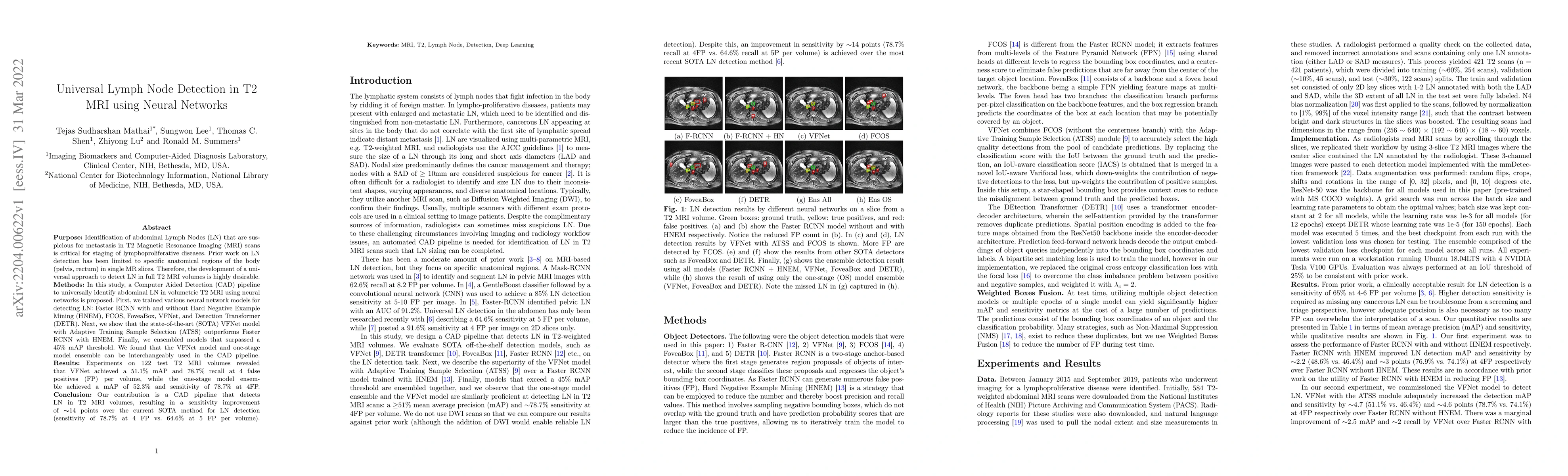
Purpose: Identification of abdominal Lymph Nodes (LN) that are suspicious for metastasis in T2 Magnetic Resonance Imaging (MRI) scans is critical for staging of lymphoproliferative diseases. Prior work on LN detection has been limited to specific anatomical regions of the body (pelvis, rectum) in single MR slices. Therefore, the development of a universal approach to detect LN in full T2 MRI volumes is highly desirable. Methods: In this study, a Computer Aided Detection (CAD) pipeline to universally identify abdominal LN in volumetric T2 MRI using neural networks is proposed. First, we trained various neural network models for detecting LN: Faster RCNN with and without Hard Negative Example Mining (HNEM), FCOS, FoveaBox, VFNet, and Detection Transformer (DETR). Next, we show that the state-of-the-art (SOTA) VFNet model with Adaptive Training Sample Selection (ATSS) outperforms Faster RCNN with HNEM. Finally, we ensembled models that surpassed a 45% mAP threshold. We found that the VFNet model and one-stage model ensemble can be interchangeably used in the CAD pipeline. Results: Experiments on 122 test T2 MRI volumes revealed that VFNet achieved a 51.1% mAP and 78.7% recall at 4 false positives (FP) per volume, while the one-stage model ensemble achieved a mAP of 52.3% and sensitivity of 78.7% at 4FP. Conclusion: Our contribution is a CAD pipeline that detects LN in T2 MRI volumes, resulting in a sensitivity improvement of $\sim$14 points over the current SOTA method for LN detection (sensitivity of 78.7% at 4 FP vs. 64.6% at 5 FP per volume).
AI Key Findings
Get AI-generated insights about this paper's methodology, results, significance, and more — seven facets brought into focus.
Impact
Paper Details
Authors
PDF Preview
Key Terms
Citation Network
Current paper (gray), citations (green), references (blue)
Display is limited for performance on very large graphs.
Discussion 0