Academic Profile
Statistics
Similar Authors
Papers on arXiv
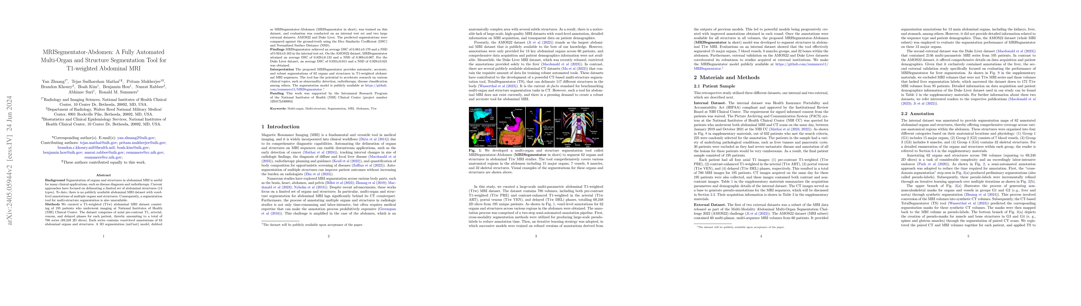
Background: Segmentation of organs and structures in abdominal MRI is useful for many clinical applications, such as disease diagnosis and radiotherapy. Current approaches have focused on delineating ...
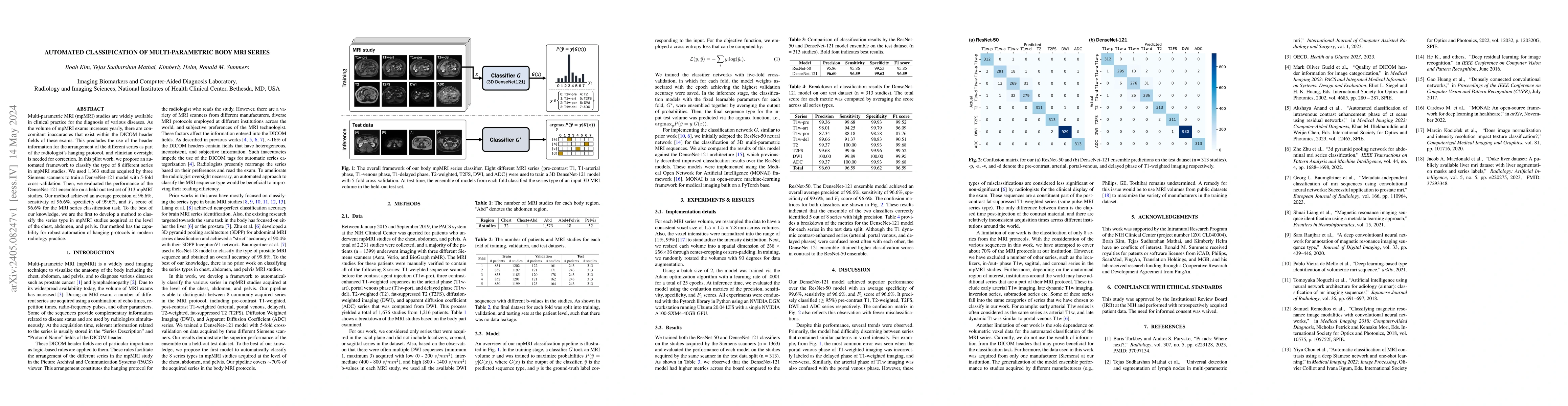
Multi-parametric MRI (mpMRI) studies are widely available in clinical practice for the diagnosis of various diseases. As the volume of mpMRI exams increases yearly, there are concomitant inaccuracie...
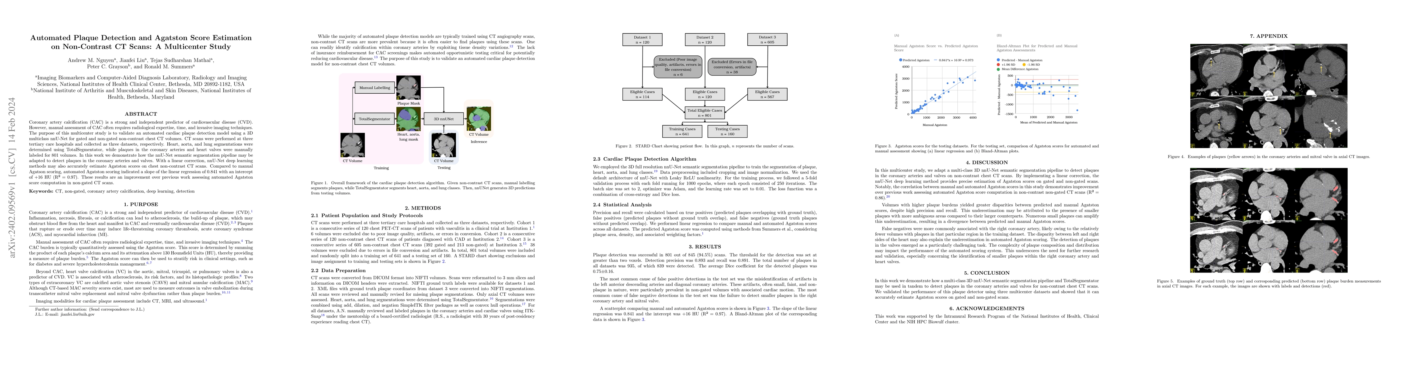
Coronary artery calcification (CAC) is a strong and independent predictor of cardiovascular disease (CVD). However, manual assessment of CAC often requires radiological expertise, time, and invasive...
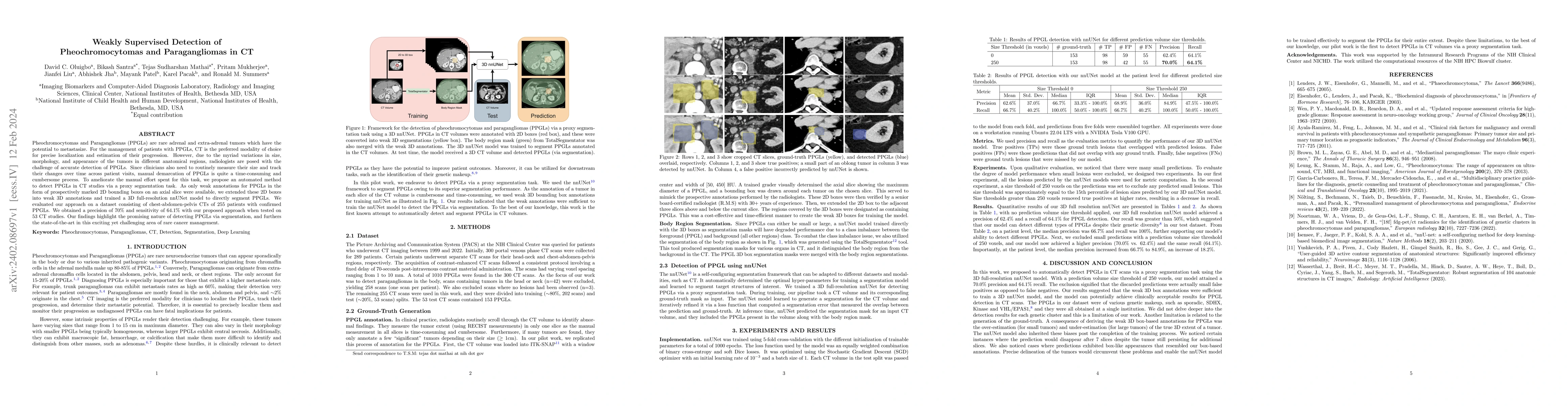
Pheochromocytomas and Paragangliomas (PPGLs) are rare adrenal and extra-adrenal tumors which have the potential to metastasize. For the management of patients with PPGLs, CT is the preferred modalit...
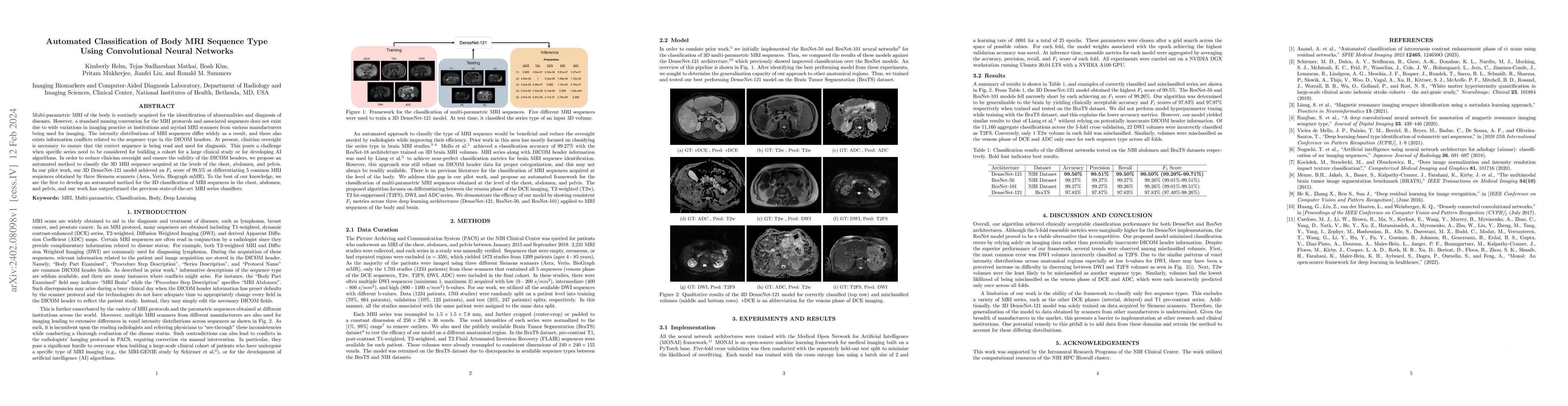
Multi-parametric MRI of the body is routinely acquired for the identification of abnormalities and diagnosis of diseases. However, a standard naming convention for the MRI protocols and associated s...
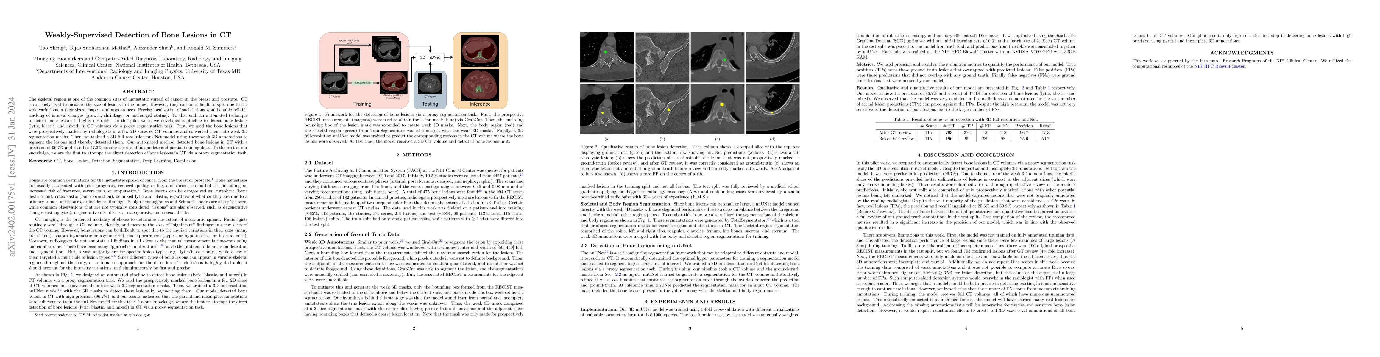
The skeletal region is one of the common sites of metastatic spread of cancer in the breast and prostate. CT is routinely used to measure the size of lesions in the bones. However, they can be diffi...
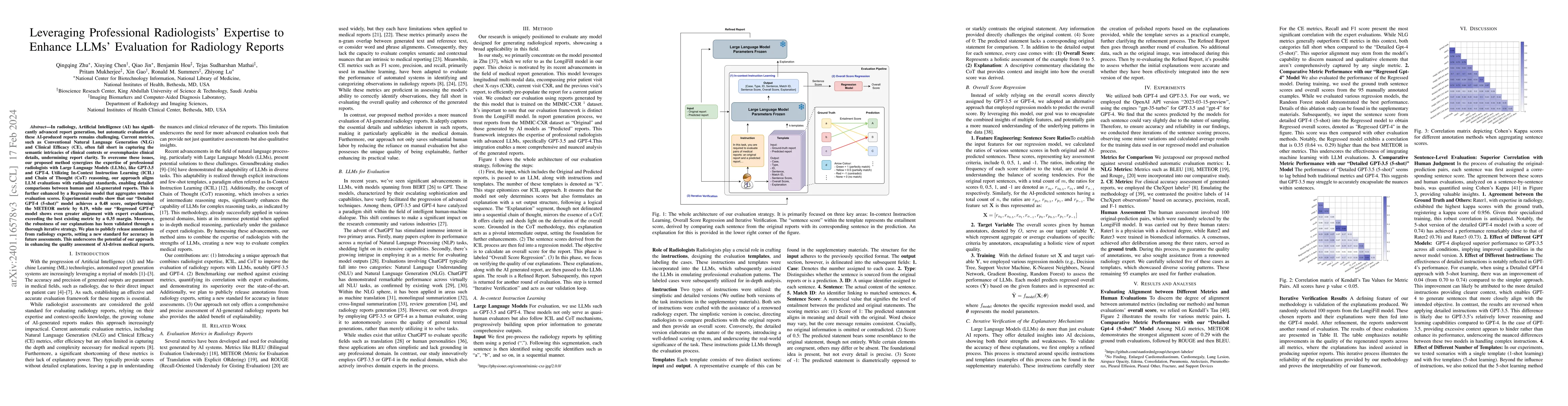
In radiology, Artificial Intelligence (AI) has significantly advanced report generation, but automatic evaluation of these AI-produced reports remains challenging. Current metrics, such as Conventio...
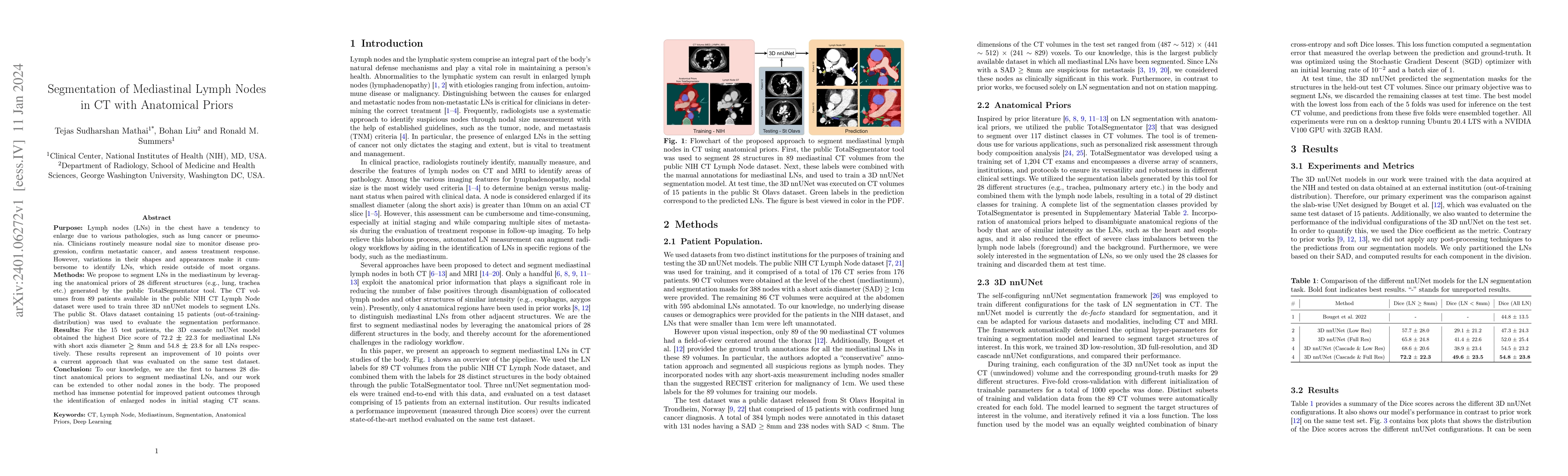
Purpose: Lymph nodes (LNs) in the chest have a tendency to enlarge due to various pathologies, such as lung cancer or pneumonia. Clinicians routinely measure nodal size to monitor disease progressio...
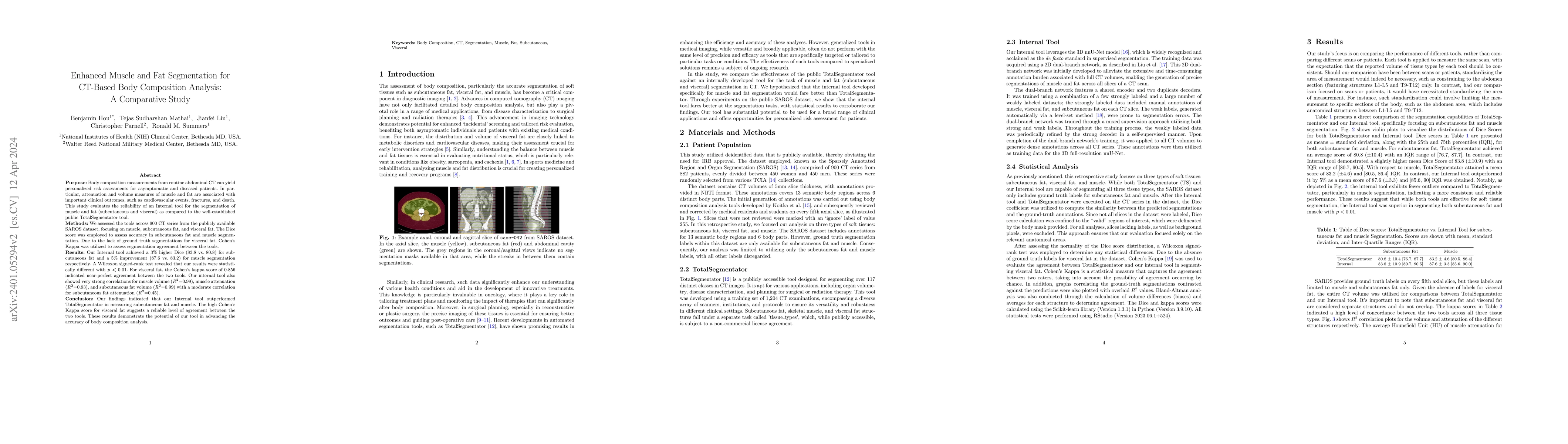
Purpose: Body composition measurements from routine abdominal CT can yield personalized risk assessments for asymptomatic and diseased patients. In particular, attenuation and volume measures of mus...
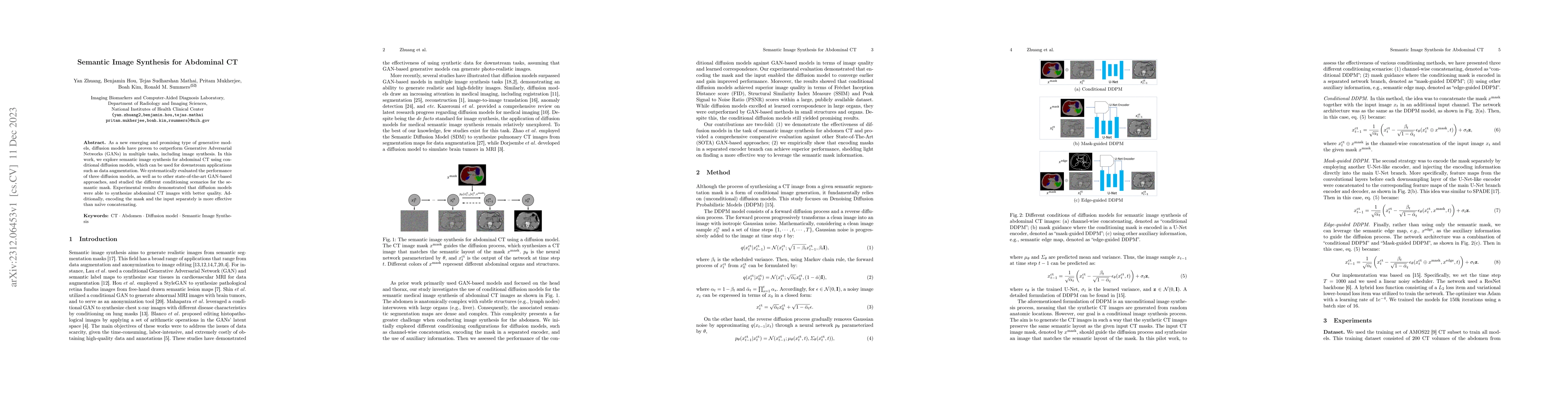
As a new emerging and promising type of generative models, diffusion models have proven to outperform Generative Adversarial Networks (GANs) in multiple tasks, including image synthesis. In this wor...
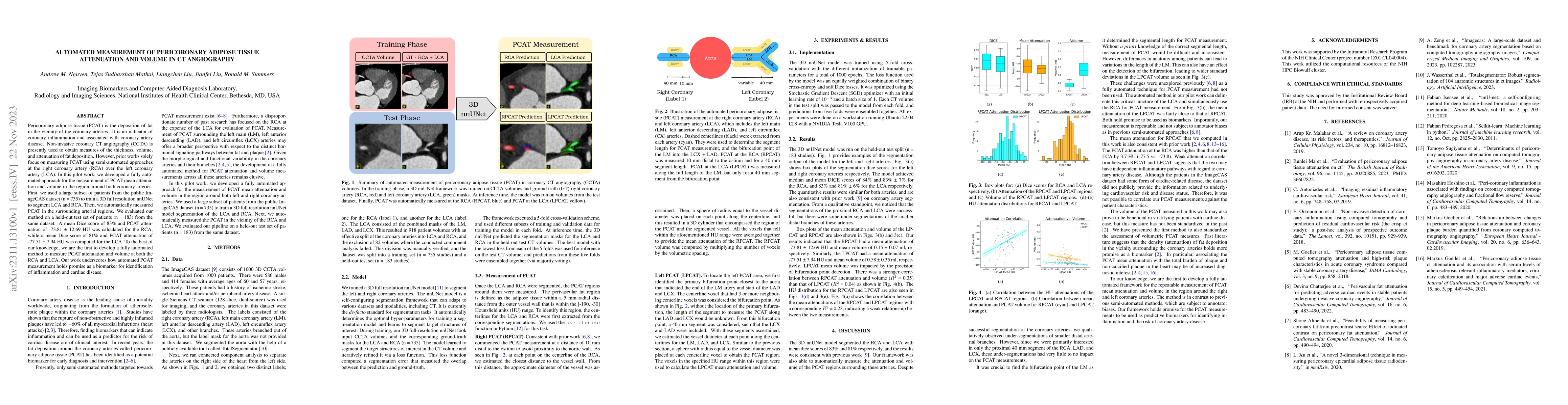
Pericoronary adipose tissue (PCAT) is the deposition of fat in the vicinity of the coronary arteries. It is an indicator of coronary inflammation and associated with coronary artery disease. Non-inv...
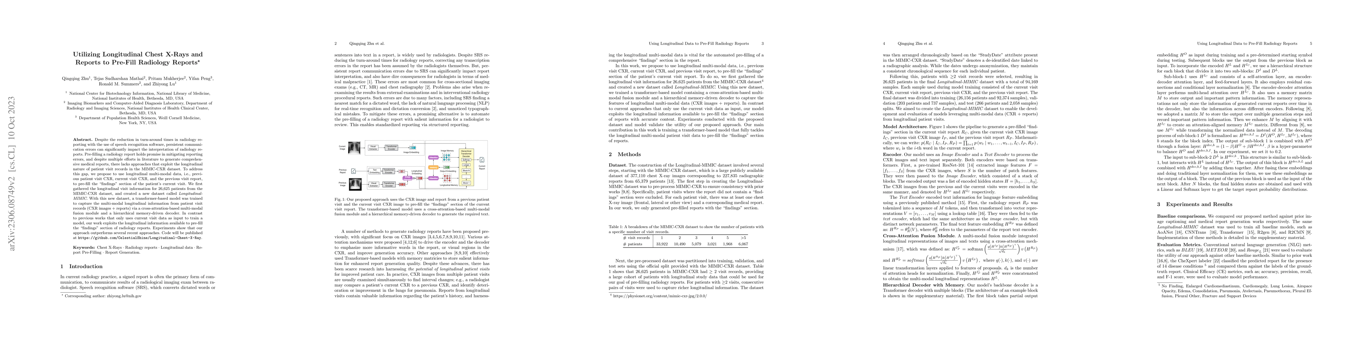
Despite the reduction in turn-around times in radiology reports with the use of speech recognition software, persistent communication errors can significantly impact the interpretation of the radiol...
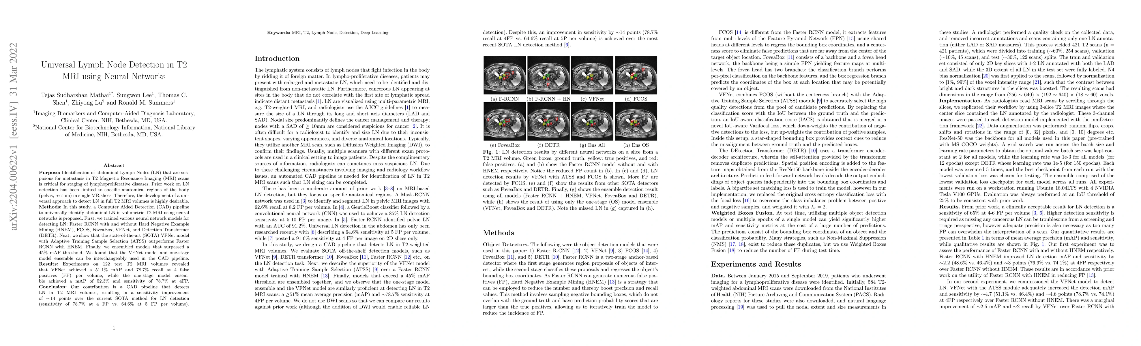
Purpose: Identification of abdominal Lymph Nodes (LN) that are suspicious for metastasis in T2 Magnetic Resonance Imaging (MRI) scans is critical for staging of lymphoproliferative diseases. Prior w...
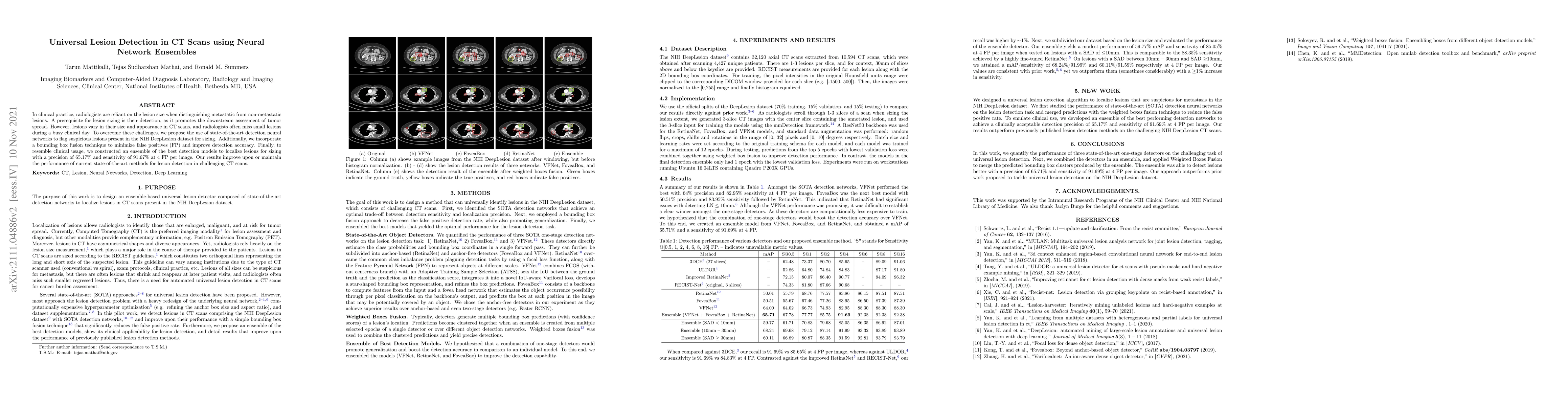
In clinical practice, radiologists are reliant on the lesion size when distinguishing metastatic from non-metastatic lesions. A prerequisite for lesion sizing is their detection, as it promotes the ...
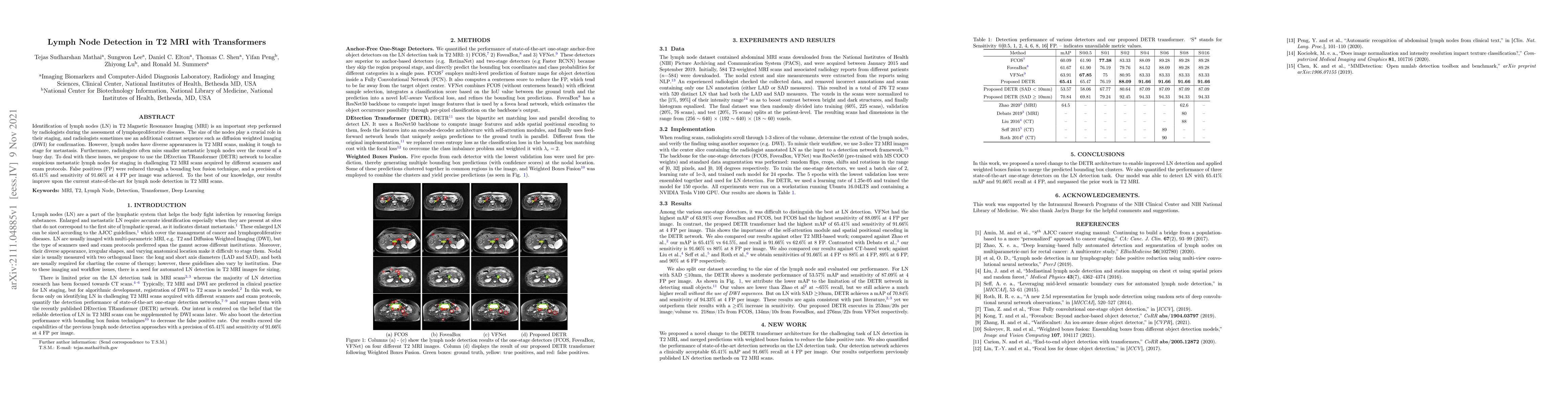
Identification of lymph nodes (LN) in T2 Magnetic Resonance Imaging (MRI) is an important step performed by radiologists during the assessment of lymphoproliferative diseases. The size of the nodes ...
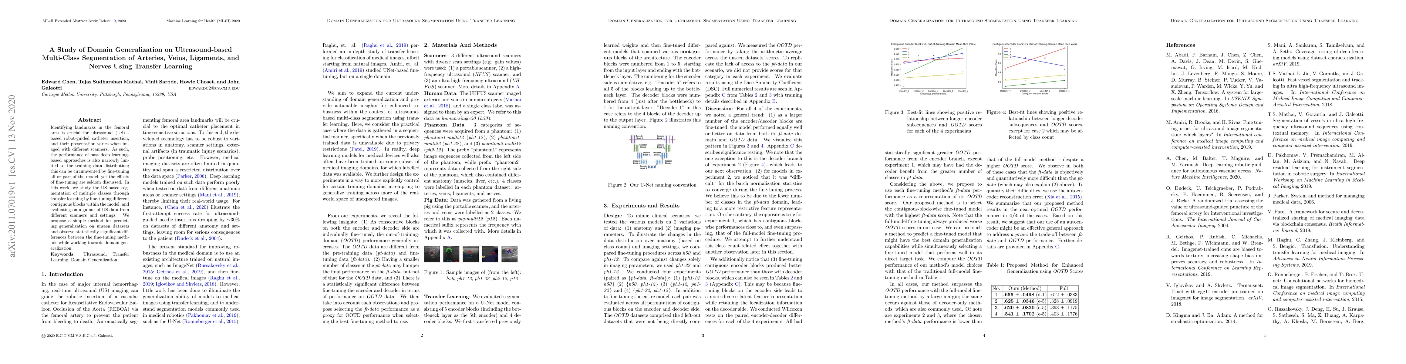
Identifying landmarks in the femoral area is crucial for ultrasound (US) -based robot-guided catheter insertion, and their presentation varies when imaged with different scanners. As such, the perfo...
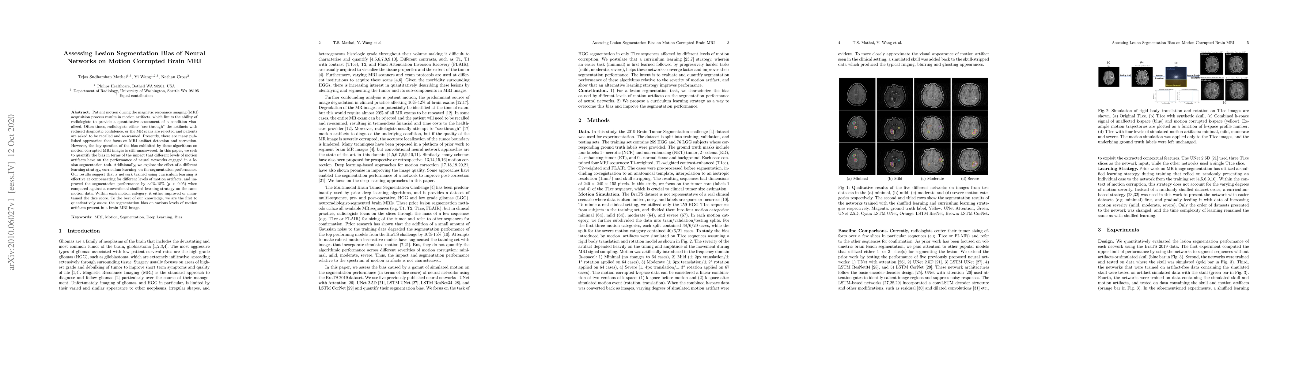
Patient motion during the magnetic resonance imaging (MRI) acquisition process results in motion artifacts, which limits the ability of radiologists to provide a quantitative assessment of a conditi...
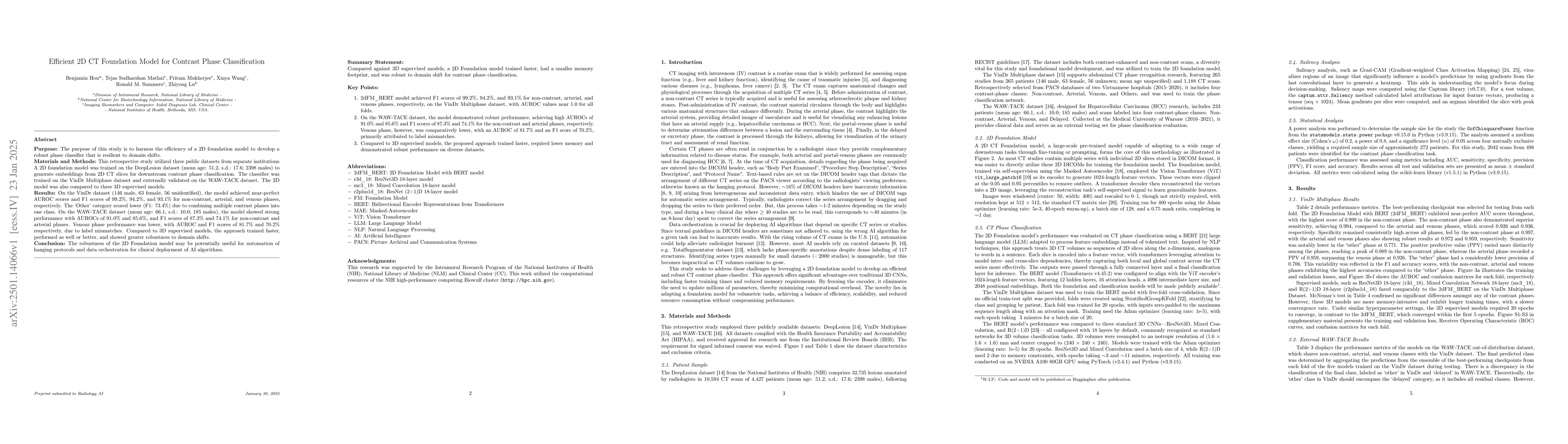
Purpose: The purpose of this study is to harness the efficiency of a 2D foundation model to develop a robust phase classifier that is resilient to domain shifts. Materials and Methods: This retrospe...
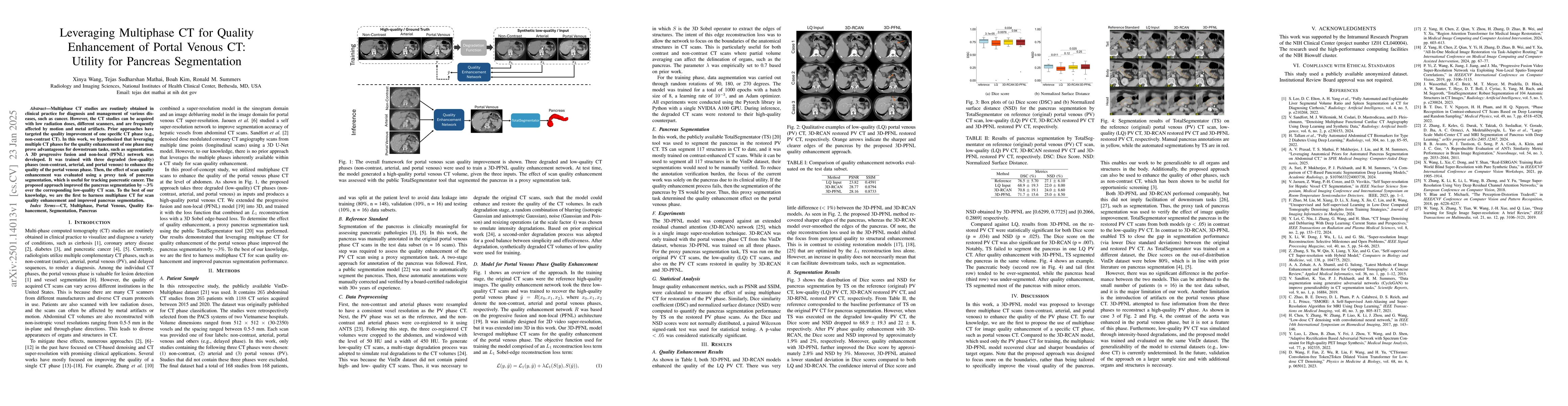
Multiphase CT studies are routinely obtained in clinical practice for diagnosis and management of various diseases, such as cancer. However, the CT studies can be acquired with low radiation doses, di...
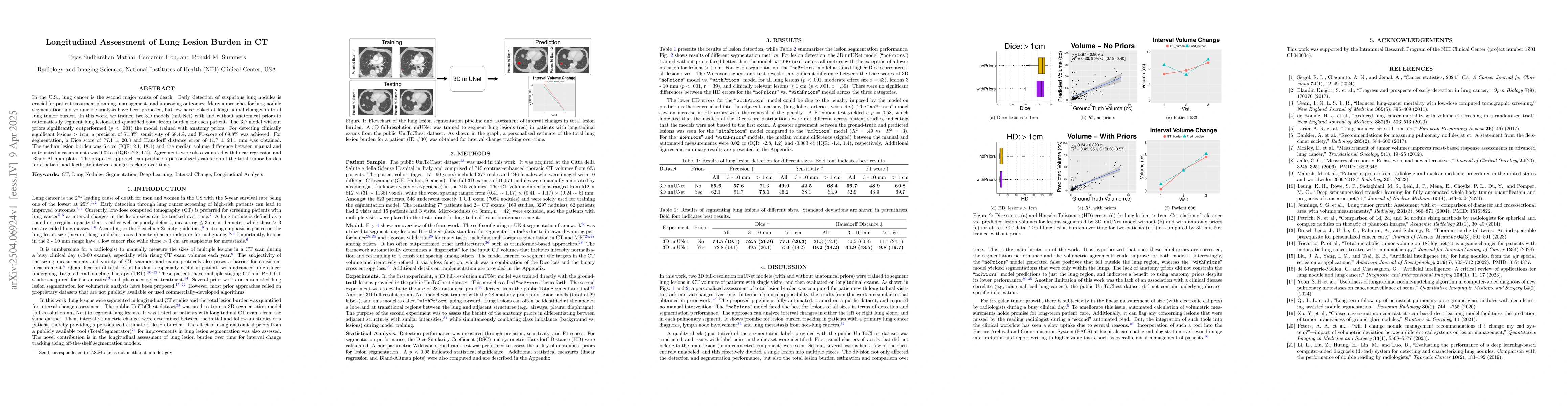
In the U.S., lung cancer is the second major cause of death. Early detection of suspicious lung nodules is crucial for patient treatment planning, management, and improving outcomes. Many approaches f...
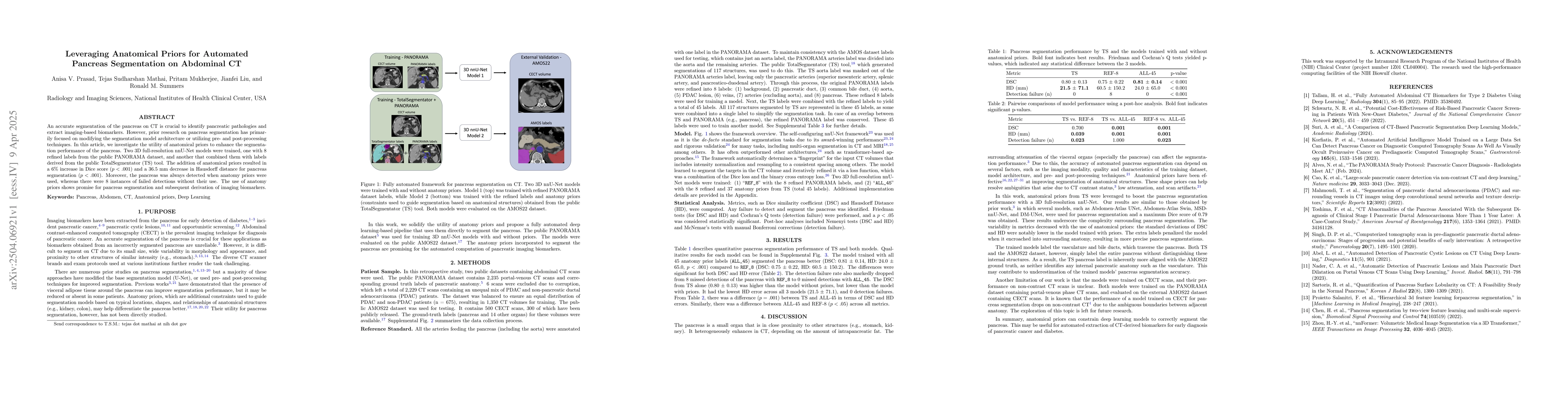
An accurate segmentation of the pancreas on CT is crucial to identify pancreatic pathologies and extract imaging-based biomarkers. However, prior research on pancreas segmentation has primarily focuse...
Radiologists routinely detect and size lesions in CT to stage cancer and assess tumor burden. To potentially aid their efforts, multiple lesion detection algorithms have been developed with a large pu...
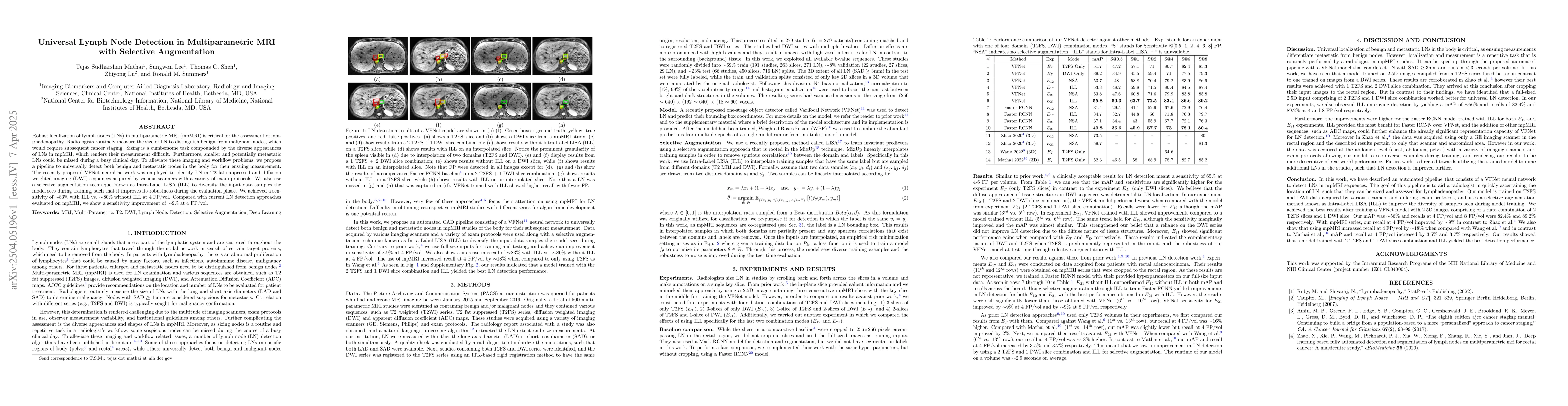
Robust localization of lymph nodes (LNs) in multiparametric MRI (mpMRI) is critical for the assessment of lymphadenopathy. Radiologists routinely measure the size of LN to distinguish benign from mali...
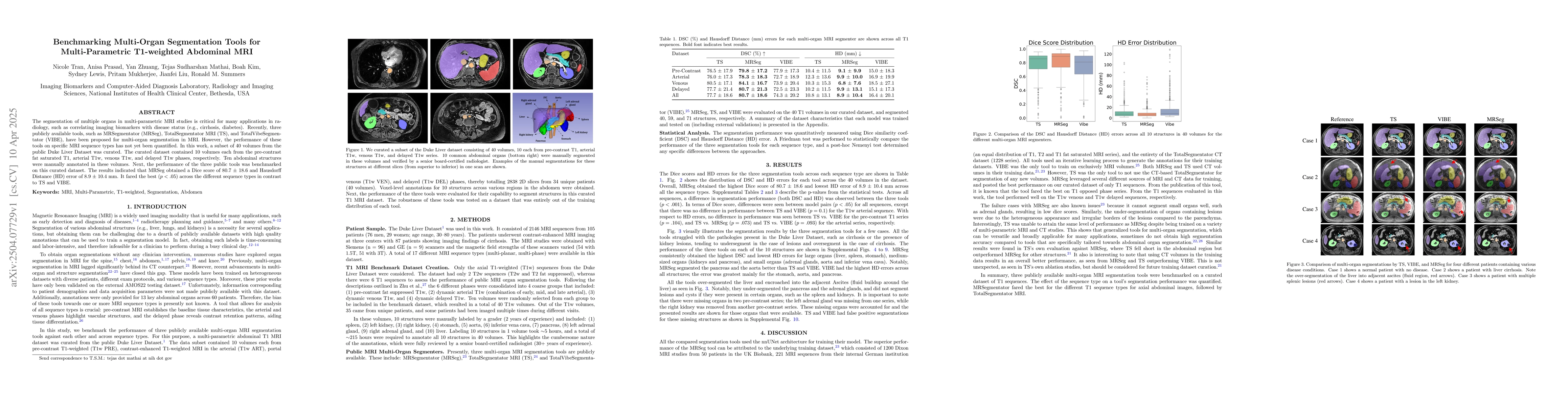
The segmentation of multiple organs in multi-parametric MRI studies is critical for many applications in radiology, such as correlating imaging biomarkers with disease status (e.g., cirrhosis, diabete...
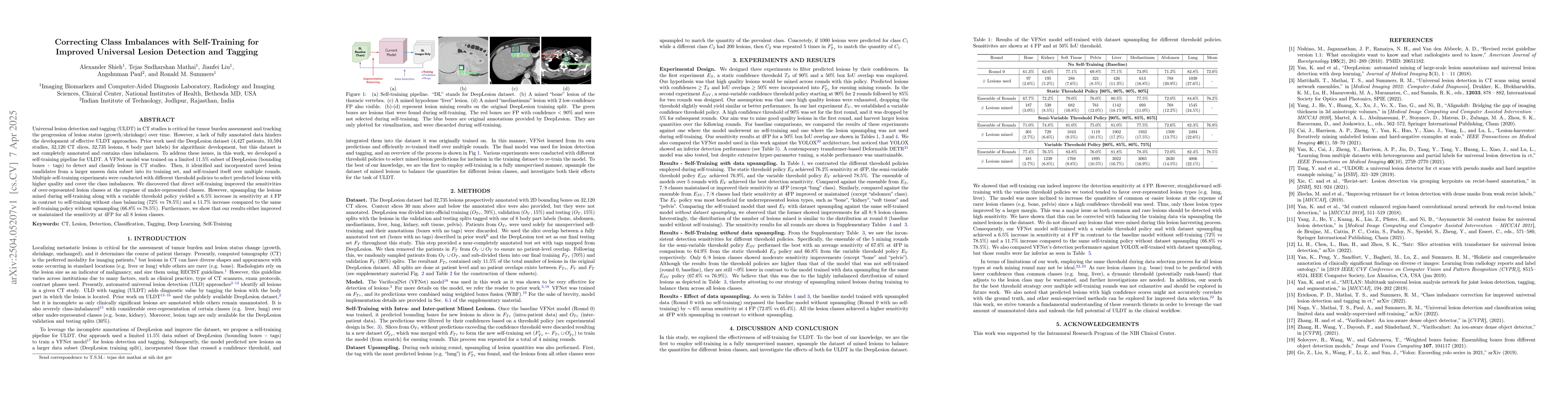
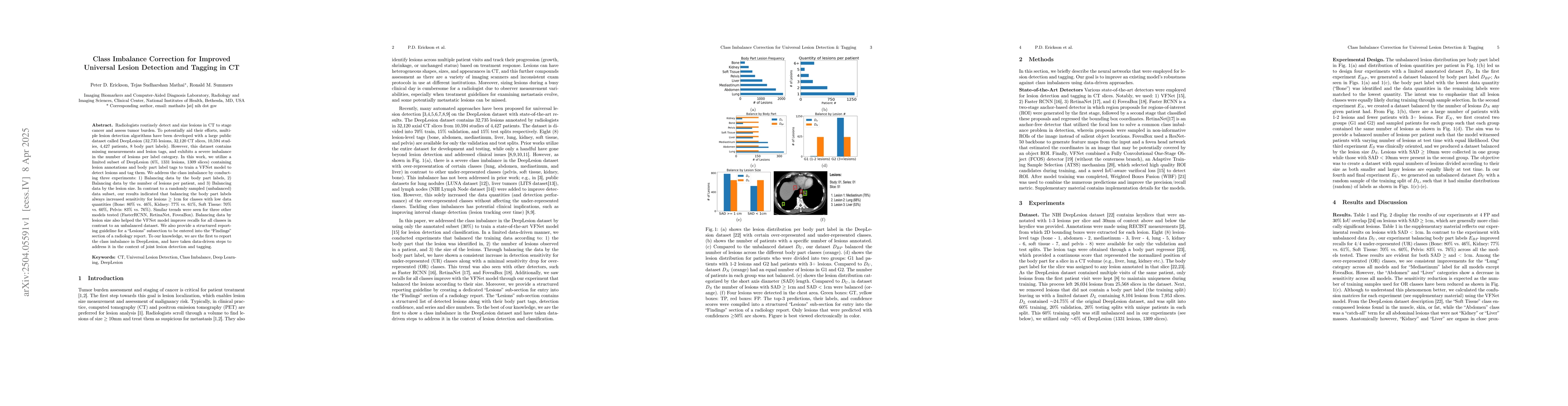
Universal lesion detection and tagging (ULDT) in CT studies is critical for tumor burden assessment and tracking the progression of lesion status (growth/shrinkage) over time. However, a lack of fully...
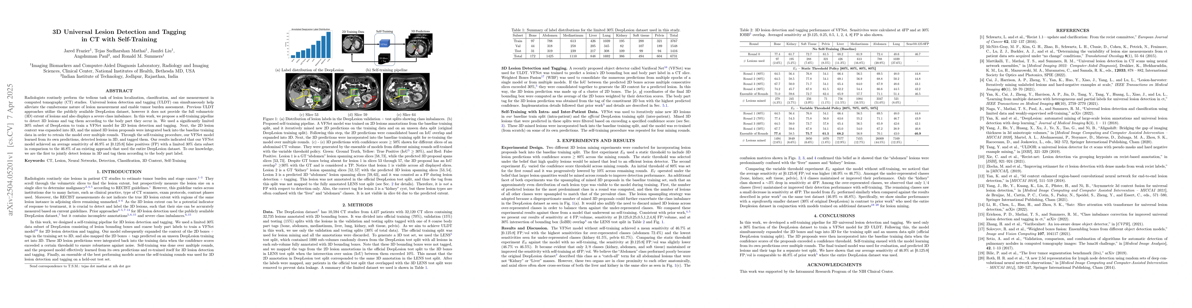
Radiologists routinely perform the tedious task of lesion localization, classification, and size measurement in computed tomography (CT) studies. Universal lesion detection and tagging (ULDT) can simu...
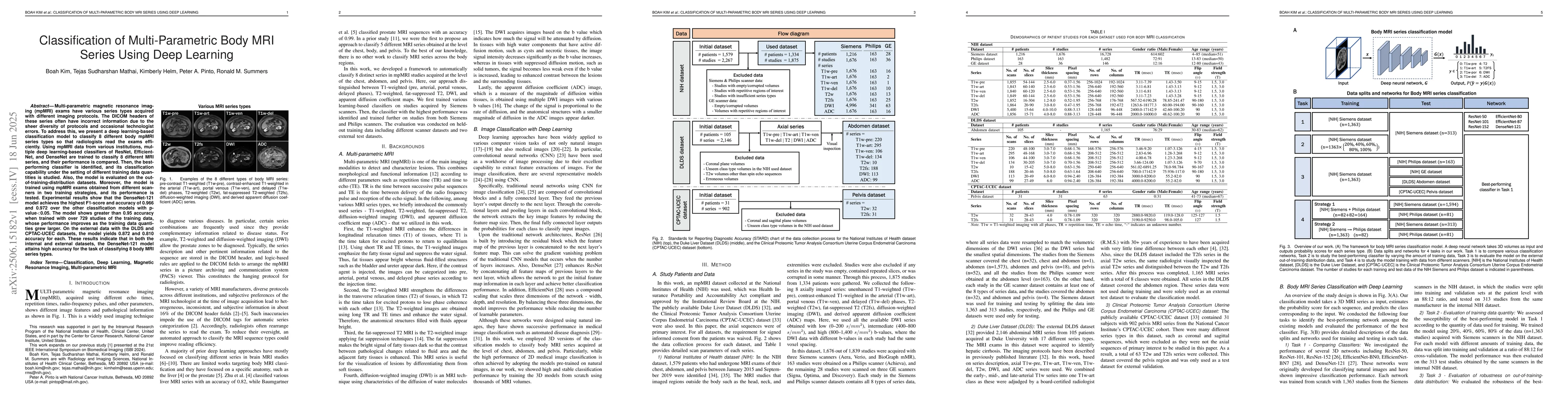
Multi-parametric magnetic resonance imaging (mpMRI) exams have various series types acquired with different imaging protocols. The DICOM headers of these series often have incorrect information due to...
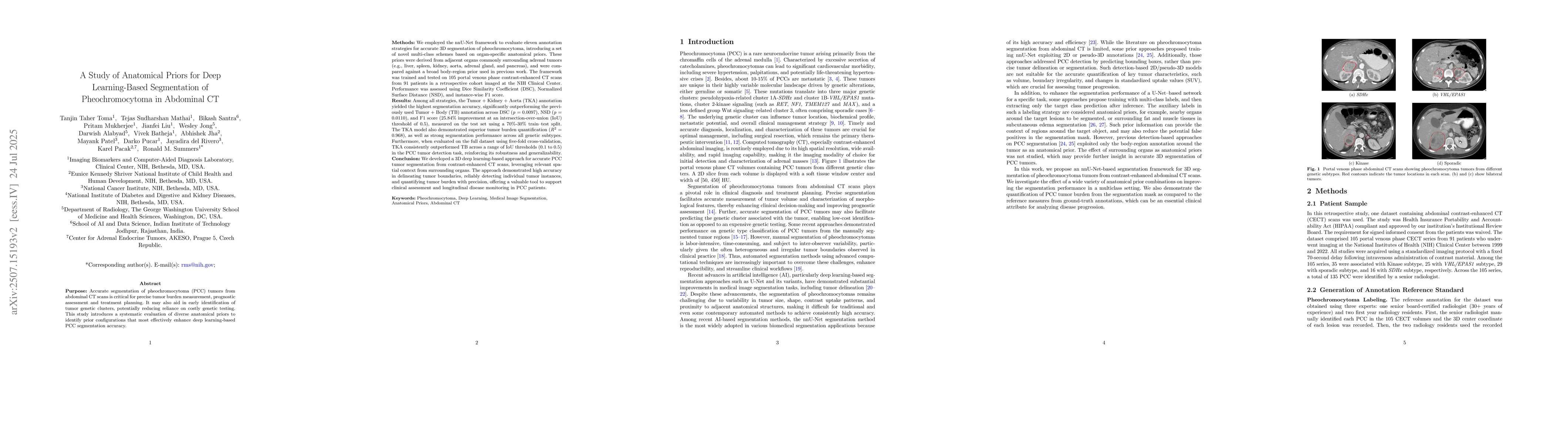
Accurate segmentation of pheochromocytoma (PCC) in abdominal CT scans is essential for tumor burden estimation, prognosis, and treatment planning. It may also help infer genetic clusters, reducing rel...
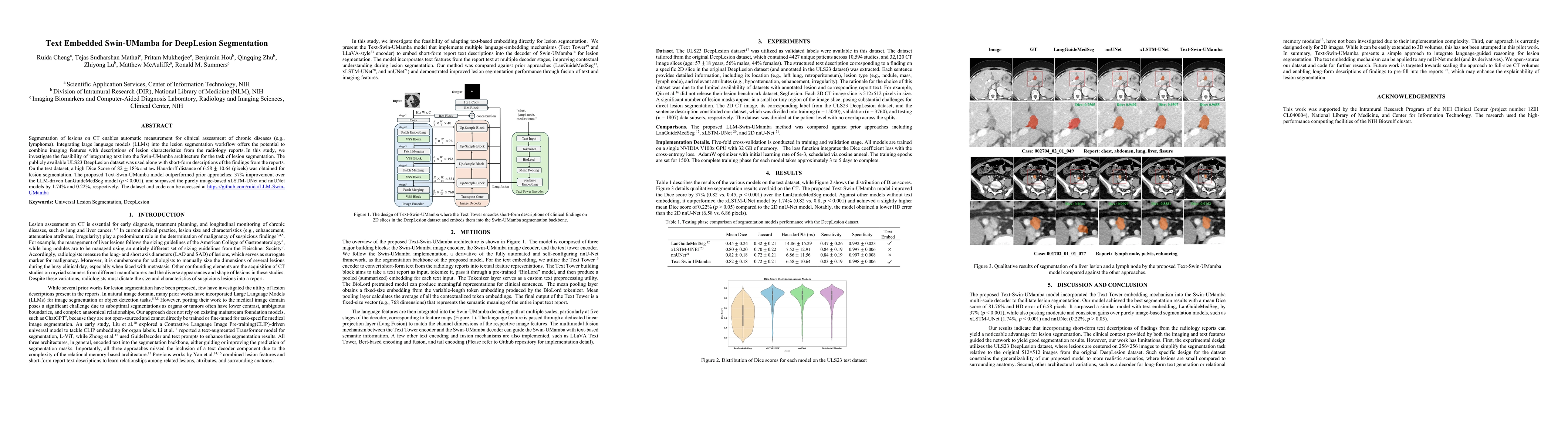
Segmentation of lesions on CT enables automatic measurement for clinical assessment of chronic diseases (e.g., lymphoma). Integrating large language models (LLMs) into the lesion segmentation workflow...
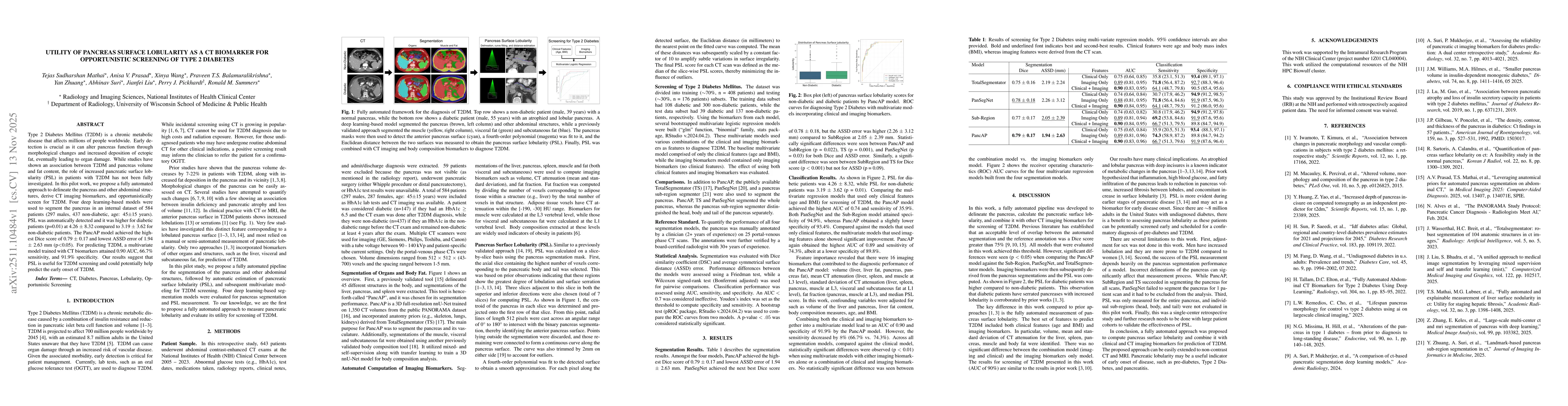
Type 2 Diabetes Mellitus (T2DM) is a chronic metabolic disease that affects millions of people worldwide. Early detection is crucial as it can alter pancreas function through morphological changes and...