Leveraging Anatomical Priors for Automated Pancreas Segmentation on Abdominal CT

Source: ArXiv

AI Quick Summary

This paper explores the integration of anatomical priors to improve automated pancreas segmentation on abdominal CT scans, achieving a 6% increase in Dice score and a significant reduction in Hausdorff distance, demonstrating the potential of anatomical knowledge to enhance segmentation accuracy.

Share: Share on X LinkedIn

Summary

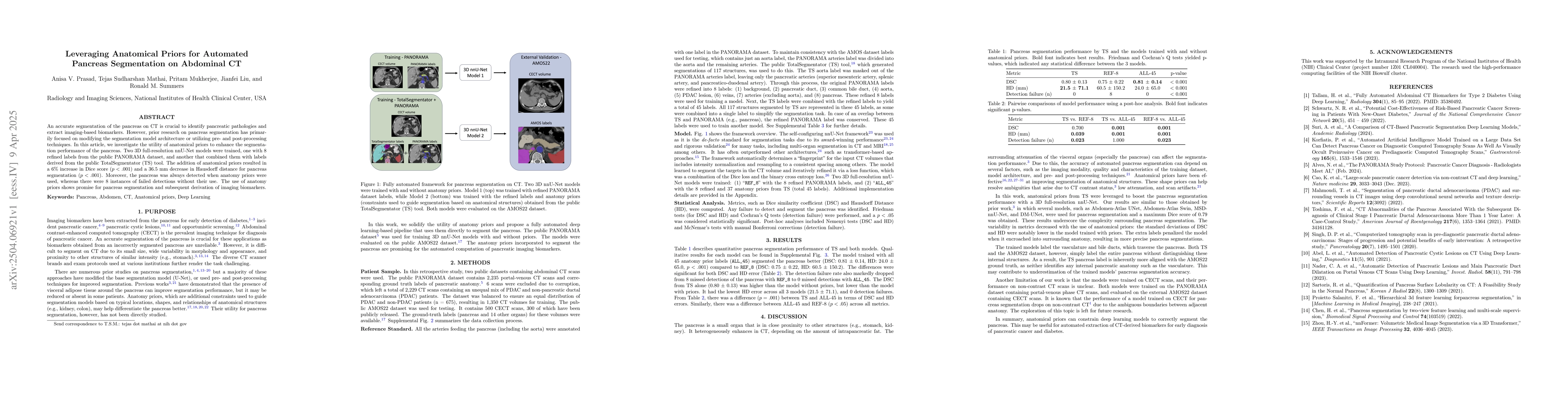
An accurate segmentation of the pancreas on CT is crucial to identify pancreatic pathologies and extract imaging-based biomarkers. However, prior research on pancreas segmentation has primarily focused on modifying the segmentation model architecture or utilizing pre- and post-processing techniques. In this article, we investigate the utility of anatomical priors to enhance the segmentation performance of the pancreas. Two 3D full-resolution nnU-Net models were trained, one with 8 refined labels from the public PANORAMA dataset, and another that combined them with labels derived from the public TotalSegmentator (TS) tool. The addition of anatomical priors resulted in a 6\% increase in Dice score ($p < .001$) and a 36.5 mm decrease in Hausdorff distance for pancreas segmentation ($p < .001$). Moreover, the pancreas was always detected when anatomy priors were used, whereas there were 8 instances of failed detections without their use. The use of anatomy priors shows promise for pancreas segmentation and subsequent derivation of imaging biomarkers.

AI Key Findings

Generated Jun 09, 2025

Methodology

This research utilized two 3D full-resolution nnU-Net models trained on refined labels from the PANORAMA dataset and combined with labels from the public TotalSegmentator (TS) tool, leveraging anatomical priors for pancreas segmentation on abdominal CT scans.

Key Results

  • Incorporation of anatomical priors increased Dice score by 6% (p < .001) and decreased Hausdorff distance by 36.5 mm (p < .001) for pancreas segmentation.
  • Pancreas detection was always successful when anatomy priors were used, whereas there were 8 instances of failed detections without their use.
  • The model trained with all 45 anatomy prior labels ('ALL_45') outperformed the model trained with only 8 refined PANORAMA labels ('REF_8') in terms of Dice similarity coefficient (DSC) and Hausdorff Distance (HD).

Significance

This study demonstrates the potential of anatomical priors in enhancing pancreas segmentation accuracy, which is crucial for identifying pancreatic pathologies and deriving imaging-based biomarkers.

Technical Contribution

The research introduced a novel approach by combining refined PANORAMA dataset labels with additional anatomical priors from the TotalSegmentator tool, enhancing pancreas segmentation using the nnU-Net framework.

Novelty

This work distinguishes itself by focusing on the utility of anatomical priors for improving pancreas segmentation, unlike previous research that primarily concentrated on modifying segmentation model architectures or employing pre- and post-processing techniques.

Limitations

  • The models were trained on contrast-enhanced CT (CECT) scans, and their performance on non-contrast CT scans remains unclear.
  • The study did not explore the impact of anatomical priors on other imaging modalities like MRI.

Future Work

  • Investigate the applicability of anatomical priors in non-contrast CT scans for pancreas segmentation.
  • Explore the use of anatomical priors in other imaging modalities, such as MRI, for organ segmentation.

Paper Details

PDF Preview

Download PDF

Citation Network

Current paper (gray), citations (green), references (blue)

Display is limited for performance on very large graphs.

Current Paper
Citations
References
Click to view
Title Authors Year Actions

Comments (0)

This website uses cookies to ensure you get the best experience. Learn more