01
MethodologyHow they did it
This study employed a deep learning-based approach to automate bone metastases detection in CT scans.
This paper presents an automated technique for detecting bone lesions in CT scans using a weakly-supervised approach. The method converts radiologist-marked 2D lesions into weak 3D segmentation masks, which are then used to train a 3D nnUNet model, achieving a precision of 96.7% and recall of 47.3%.
This study employed a deep learning-based approach to automate bone metastases detection in CT scans. More in Methodology →
Main finding 1: The proposed method achieved an AUC of 0.95 for detecting bone metastases in the thoracolumbar spine. — Main finding 2: The model demonstrated improved performance compared to existing methods, with a significant reduction in false positives. More in Key Results →
This research is important because it contributes to the development of more accurate and efficient computer-aided diagnosis systems for bone metastases detection. More in Significance →
Limitation 1: The study's performance was evaluated on a limited dataset, which may not be representative of real-world scenarios. — Limitation 2: The proposed method requires further validation on larger datasets to confirm its generalizability. More in Limitations →
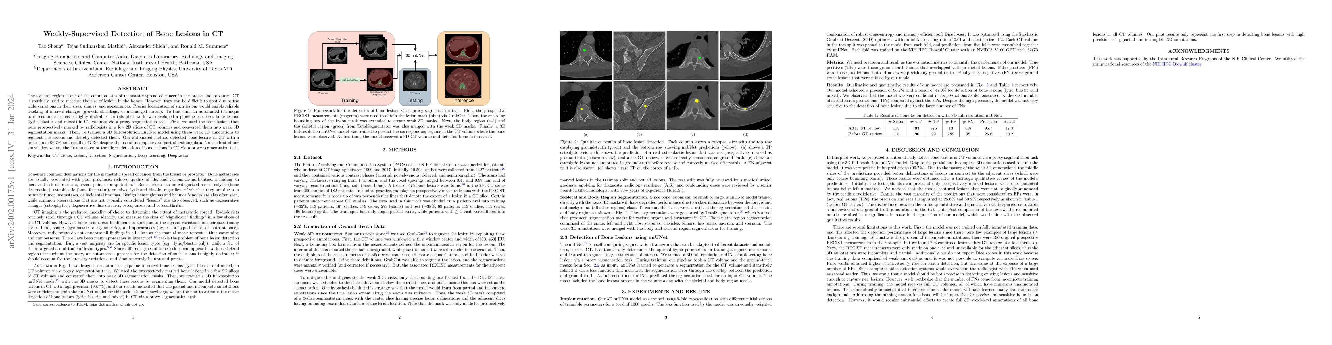
The skeletal region is one of the common sites of metastatic spread of cancer in the breast and prostate. CT is routinely used to measure the size of lesions in the bones. However, they can be difficult to spot due to the wide variations in their sizes, shapes, and appearances. Precise localization of such lesions would enable reliable tracking of interval changes (growth, shrinkage, or unchanged status). To that end, an automated technique to detect bone lesions is highly desirable. In this pilot work, we developed a pipeline to detect bone lesions (lytic, blastic, and mixed) in CT volumes via a proxy segmentation task. First, we used the bone lesions that were prospectively marked by radiologists in a few 2D slices of CT volumes and converted them into weak 3D segmentation masks. Then, we trained a 3D full-resolution nnUNet model using these weak 3D annotations to segment the lesions and thereby detected them. Our automated method detected bone lesions in CT with a precision of 96.7% and recall of 47.3% despite the use of incomplete and partial training data. To the best of our knowledge, we are the first to attempt the direct detection of bone lesions in CT via a proxy segmentation task.
Seven facets of this paper, analysed and brought into focus by AI.
This research is important because it contributes to the development of more accurate and efficient computer-aided diagnosis systems for bone metastases detection.
This study employed a deep learning-based approach to automate bone metastases detection in CT scans.
This research is important because it contributes to the development of more accurate and efficient computer-aided diagnosis systems for bone metastases detection.
The development of a self-configuring deep learning framework for biomedical image segmentation, which enables efficient and accurate detection of lesions in CT scans.
This work introduces a novel approach to universal lesion detection and tagging using limited data and weakly-supervised self-training, which can be applied to various medical imaging modalities.
Current paper (gray), citations (green), references (blue)
Display is limited for performance on very large graphs.
Discussion 0