ArxivLens

Quick Summary:

This paper introduces a method for improving quality control in molecular optical sectioning microscopy by using concentration-encoded effective photons to standardize pixel measurements. The proposed tool enables objective comparisons of imaging performance across different microscopy systems and settings, facilitating quantitative imaging in live specimens.

arXiv Id: 2307.04670
Date Published: 2023-07-11
Date Updated: 2023-07-11

Key Terms (TF-IDF):

quantitative (0.234)
microscopy (0.223)
tool (0.221)
imaging (0.185)
molecular (0.179)
measurement (0.170)
fluorescence (0.152)
illumination (0.152)
labeling (0.152)
indispensable (0.149)
descriptions (0.143)
comparisons (0.140)
signal noise (0.139)
modalities (0.138)
frames (0.137)
acquired (0.137)
acquisition (0.136)
attractive (0.133)
numerous (0.130)
qualitative (0.129)
License: http://creativecommons.org/licenses/by/4.0/

Summary:

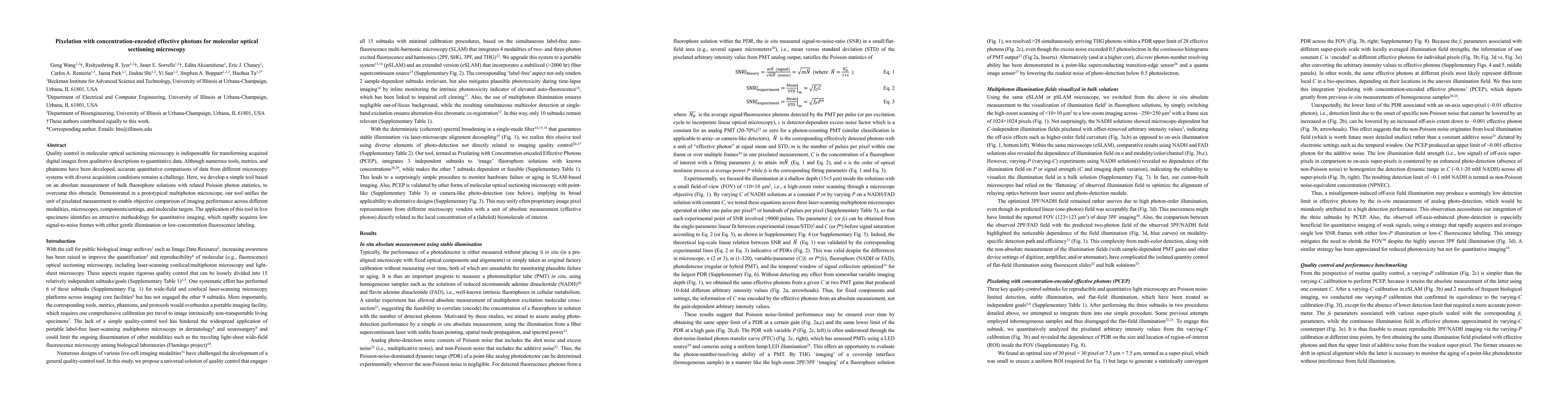
Quality control in molecular optical sectioning microscopy is indispensable for transforming acquired digital images from qualitative descriptions to quantitative data. Although numerous tools, metrics, and phantoms have been developed, accurate quantitative comparisons of data from different microscopy systems with diverse acquisition conditions remains a challenge. Here, we develop a simple tool based on an absolute measurement of bulk fluorophore solutions with related Poisson photon statistics, to overcome this obstacle. Demonstrated in a prototypical multiphoton microscope, our tool unifies the unit of pixelated measurement to enable objective comparison of imaging performance across different modalities, microscopes, components/settings, and molecular targets. The application of this tool in live specimens identifies an attractive methodology for quantitative imaging, which rapidly acquires low signal-to-noise frames with either gentle illumination or low-concentration fluorescence labeling.

Citation & Reference Network Visualization

This graph shows how this paper is connected to other research. The current paper is shown in dark gray, papers that cite it are in green, and papers it references are in blue. You can:

Current Paper
Papers citing this work (Citations)
Papers cited by this work (References)

• Hover over any node to see the paper title

• Click a node to view that paper's details

• Drag nodes to rearrange the graph

• Use the zoom controls (bottom right) to zoom in/out

Citations and References

Title Authors Year Actions
Analog multiplexing of a laser clock and computational photon counting for fast fluorescence lifetime imaging microscopy
Rishyashring R. Iyer, Janet E. Sorrells, Kevin Tan, Lingxiao Yang, Geng Wang, Haohua Tu, Stephen A. Boppart
2024
View Paper
Supercontinuum intrinsic fluorescence imaging heralds ‘free view’ of living systems
Geng Wang, Lianhuang Li, Xiaoxia Liao, Shu Wang, Jennifer Mitchell, R. “. Rabel, Shirui Luo, Jindou Shi, Janet E. Sorrells, Rishyashring R. Iyer, Edita Aksamitiene, Carlos A Renteria, E. Chaney, Derek J. Milner, Matthew B. Wheeler, Martha U. Gillette, Alexander G. Schwing, Jianxin Chen, Haohua Tu
2024
View Paper
Gentle label-free nonlinear optical imaging relaxes linear-absorption-mediated triplet
Geng Wang, Lianhuang Li, Janet E. Sorrells, Jianxin Chen, H. Tu
2023
View Paper

Similar Papers

Smartphone-based Optical Sectioning (SOS) Microscopy with A Telecentric Design for Fluorescence Imaging

Yu Chen, Mingliang Pan, David Griffin, Ziao Jiao, Khadija Yousaf, Daniel Doveiko, Michelle Maclean, David Day Uei Lia

Published: 2023-10-04

Structured Detection for Simultaneous Super-Resolution and Optical Sectioning in Laser Scanning Microscopy

Alessandro Zunino, Giacomo Garrè, Eleonora Perego, Sabrina Zappone, Mattia Donato, Giuseppe Vicidomini

Published: 2024-06-19

Comments (0)

This website uses cookies to ensure you get the best experience. Learn more