ArxivLens

Quick Summary:

This paper introduces Laser Light Sheet based Fluorescence Microscopy (LSFM) and its implementations, including Orthogonal-Plane Fluorescence Optical Sectioning (OPFOS), for non-destructive, high-resolution optical sectioning of biomedical specimens. It discusses the techniques' ability to visualize three-dimensional tissue details and their applications in biomedical research.

arXiv Id: 1106.3162
Comments: 19 pages, 10 figures, to be published in Anatomical Research International (Hindawi)
Date Published: 2011-06-17
Date Updated: 2011-06-17

Key Terms (TF-IDF):

fluorescence (0.352)
sheet (0.279)
microscopy (0.246)
laser (0.221)
optical (0.213)
hr (0.168)
sciences (0.168)
light (0.165)
recorded (0.157)
originating (0.152)
virtual (0.147)
camera (0.145)
emerging (0.144)
applicability (0.144)
briefly (0.142)
life (0.140)
create (0.137)
orthogonal (0.134)
methods (0.133)
cross section (0.133)
License: http://creativecommons.org/licenses/by-nc-sa/3.0/

Summary:

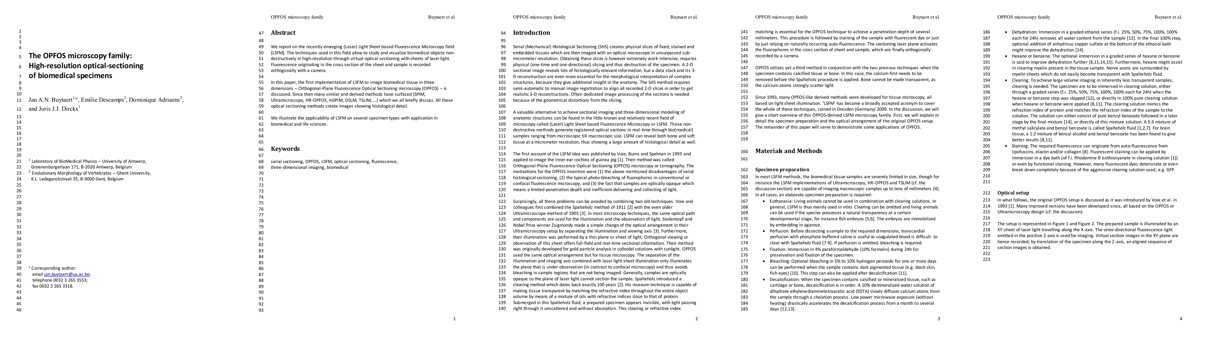
We report on the recently emerging (Laser) Light Sheet based Fluorescence Microscopy field (LSFM). The techniques used in this field allow to study and visualize biomedical objects non-destructively in high-resolution through virtual optical sectioning with sheets of laser light. Fluorescence originating in the cross section of the sheet and sample is recorded orthogonally with a camera. In this paper, the first implementation of LSFM to image biomedical tissue in three dimensions - Orthogonal-Plane Fluorescence Optical Sectioning microscopy (OPFOS) - is discussed. Since then many similar and derived methods have surfaced (SPIM, Ultramicroscopy, HR-OPFOS, mSPIM, DSLM, TSLIM...) which we all briefly discuss. All these optical sectioning methods create images showing histological detail. We illustrate the applicability of LSFM on several specimen types with application in biomedical and life sciences.

Citation & Reference Network Visualization

This graph shows how this paper is connected to other research. The current paper is shown in dark gray, papers that cite it are in green, and papers it references are in blue. You can:

Current Paper
Papers citing this work (Citations)
Papers cited by this work (References)

• Hover over any node to see the paper title

• Click a node to view that paper's details

• Drag nodes to rearrange the graph

• Use the zoom controls (bottom right) to zoom in/out

Citations and References

Title Authors Year Actions
A qualitative comparison of 3D visualization in Xenopus laevis using a traditional method and a non-destructive method
E. Descamps, J. Buytaert, B. Kegel, J. Dirckx, D. Adriaens
2024
View Paper
Lightsheet Microscopy
Laura Dyer, A. Parker, Keanu Paphiti, J. Sanderson
2022
View Paper
Light-sheet mesoscopy with the Mesolens provides fast sub-cellular resolution imaging throughout large tissue volumes
Eliana Battistella, J. Schniete, Katrina Wesencraft, J. Quintana, G. McConnell
2022
View Paper
Micro-CT imaging of Thiel-embalmed and iodine-stained human temporal bone for 3D modeling
Sebastian Halm, D. Haberthür, Elisabeth Eppler, V. Djonov, A. Arnold
2020
View Paper
Neural defects caused by total and Wnt1-Cre mediated ablation of p120ctn in mice
T. Pieters, E. Sanders, Huiyu Tian, J. van Hengel, F. van Roy
2020
View Paper
Imaging of the immune system – towards a subcellular and molecular understanding
Lai Wen, Zhichao Fan, Z. Mikulski, K. Ley
2020
View Paper
Soft tissue discrimination with contrast agents using micro-CT scanning
E. Descamps, Alicja Sochacka, B. Kegel, D. Loo, L. Hoorebeke, D. Adriaens
2020
View Paper
Endogenous erythropoietin signaling regulates migration and laminar positioning of upper-layer neurons in the developing neocortex
P. Constanthin, A. Contestabile, V. Petrenko, C. Quairiaux, P. Salmon, P. Hüppi, J. Kiss
2020
View Paper
Characterizing the size of the target region for atraumatic opening of the cochlea through the facial recess
T. Rau, D. Kreul, J. Lexow, S. Hügl, M. G. Zuniga, T. Lenarz, O. Majdani
2019
View Paper
Multiscale light-sheet for rapid imaging of cardiopulmonary system.
Yichen Ding, Jianguo Ma, Adam D. Langenbacher, K. Baek, Juhyun Lee, Chih-Chiang Chang, J. Hsu, Rajan P. Kulkarni, J. Belperio, W. Shi, Sara Ranjbarvaziri, R. Ardehali, Y. Tintut, L. Demer, Jau-Nian Chen, Peng Fei, R. Packard, T. Hsiai
2018
View Paper
The light-sheet microscopy revolution
J. Girkin, Mariana T. Carvalho
2018
View Paper
Light-Sheet Imaging to Elucidate Cardiovascular Injury and Repair
Yichen Ding, Juhyun Lee, J. Hsu, Chih-Chiang Chang, K. Baek, Sara Ranjbarvaziri, R. Ardehali, R. Packard, T. Hsiai
2018
View Paper
Integrating light-sheet imaging with virtual reality to recapitulate developmental cardiac mechanics.
Yichen Ding, Arash Abiri, Parinaz Abiri, Shuoran Li, Chih-Chiang Chang, K. Baek, J. Hsu, Elias Sideris, Yilei Li, Juhyun Lee, T. Segura, T. Nguyen, Alexander Bui, René R. Sevag Packard, Peng Fei, T. Hsiai
2017
View Paper
Improved middle-ear soft-tissue visualization using synchrotron radiation phase-contrast imaging
M. Elfarnawany, S. A. Rohani, Soroush Ghomashchi, D. Allen, N. Zhu, S. Agrawal, H. Ladak
2017
View Paper
Light-sheet fluorescence imaging to localize cardiac lineage and protein distribution
Yichen Ding, Juhyun Lee, Jianguo Ma, Kevin Sung, Tomohiro Yokota, Neha Singh, Mojdeh Dooraghi, Parinaz Abiri, Yibin Wang, Rajan P. Kulkarni, A. Nakano, T. Nguyen, Peng Fei, T. Hsiai
2017
View Paper
Advancing ovarian folliculometry with selective plane illumination microscopy
Hsiao-Chun Amy Lin, R. Dutta, S. Mandal, A. Kind, A. Schnieke, D. Razansky
2016
View Paper
Determination of the mastoid surface area and volume based on micro-CT scanning of human temporal bones. Geometrical parameters depend on scanning resolutions
O. Cros, H. Knutsson, M. Andersson, Elin Pawels, M. Borga, M. Gaihede
2016
View Paper
Visualization, measurement and modelling of the cochlea using rotating midmodiolar slice planes
G. Lexow, Daniel Schurzig, N. Gellrich, T. Lenarz, O. Majdani, T. Rau
2016
View Paper
High Fidelity Bioelectric Modelling of the Implanted Cochlea
Paul Wong
2015
View Paper
Light Sheet Fluorescence Microscopy (LSFM)
Michael W. Adams, Andrew F. Loftus, Sarah E. Dunn, Matthew S. Joens, James A.J. Fitzpatrick
2015
View Paper
Volume Shrinkage of Bone, Brain and Muscle Tissue in Sample Preparation for Micro-CT and Light Sheet Fluorescence Microscopy (LSFM)
J. Buytaert, J. Goyens, D. De Greef, P. Aerts, J. Dirckx
2014
View Paper
Micro-channels in the mastoid anatomy. Indications of a separate blood supply of the air cell system mucosa by micro-CT scanning
O. Cros, M. Borga, E. Pauwels, J. Dirckx, M. Gaihede
2013
View Paper
Three-dimensional histological specimen preparation for accurate imaging and spatial reconstruction of the middle and inner ear
T. Rau, W. Würfel, T. Lenarz, O. Majdani
2013
View Paper
MicroCT versus sTSLIM 3D Imaging of the Mouse Cochlea
J. Buytaert, Shane B. Johnson, M. Dierick, Wasil H. M. Salih, P. Santi
2013
View Paper
Realistic 3D Computer Model of the Gerbil Middle Ear, Featuring Accurate Morphology of Bone and Soft Tissue Structures
J. Buytaert, Wasil H. M. Salih, M. Dierick, P. Jacobs, J. Dirckx
2011
View Paper
Light-Sheet Fluorescence Microscopy
B. Masters
2020
View Paper
Prototyping an Open Source Single Plane Illumination Microscope for 3 D Histology
Joshua A. Berko
2018
View Paper
Light Sheet Fluorescence Microscopy Applications for Multicellular Systems
J. Colombelli, Corinne Lorenzo
2014
View Paper
Optimierung der Insertion von Cochlea-Implantaten unter Berücksichtigung des Verformungsverhaltens der Elektrodenträger: mechatronik-gestützte, minimal traumatische CI-Chirurgie
T. Rau, Berichte aus der Biomechatronik, H. Witte
2014
View Paper
Deposited in DRO : 18 April 2018 Version of attached
Achim von Arnims, C. Nitschke
2014
View Paper

Similar Papers

Super-resolution of biomedical volumes with 2D supervision

Honglak Lee, Cheng Jiang, Yiwei Lyu, Yufeng Zhang, Xinhai Hou, Akhil Kondepudi, Asadur Chowdury, Alexander Gedeon, Eric Landgraf, Todd Hollon

Published: 2024-04-16

Structured Detection for Simultaneous Super-Resolution and Optical Sectioning in Laser Scanning Microscopy

Alessandro Zunino, Giacomo Garrè, Eleonora Perego, Sabrina Zappone, Mattia Donato, Giuseppe Vicidomini

Published: 2024-06-19

Smartphone-based Optical Sectioning (SOS) Microscopy with A Telecentric Design for Fluorescence Imaging

Yu Chen, Mingliang Pan, David Griffin, Ziao Jiao, Khadija Yousaf, Daniel Doveiko, Michelle Maclean, David Day Uei Lia

Published: 2023-10-04

Pixelation with concentration-encoded effective photons for molecular optical sectioning microscopy

Yi Sun, Geng Wang, Jaena Park, Jindou Shi, Rishyashring R. Iyer, Janet E. Sorrells, Haohua Tu, Edita Aksamitiene, Eric J. Chaney, Carlos A. Renteria, Stephen A. Boppart

Published: 2023-07-11

Comments (0)

This website uses cookies to ensure you get the best experience. Learn more