Smartphone-based Optical Sectioning (SOS) Microscopy with A Telecentric Design for Fluorescence Imaging
Publication
Metrics
AI Quick Summary
This paper proposes a low-cost Smartphone-based Optical Sectioning (SOS) microscope for fluorescence imaging using a telecentric design and the HiLo technique. The SOS integrates a smartphone for illumination and imaging, achieving high contrast fluorescent imaging and optical sectioning with an axial resolution of 11.7 μm.
Paper Preview
Abstract
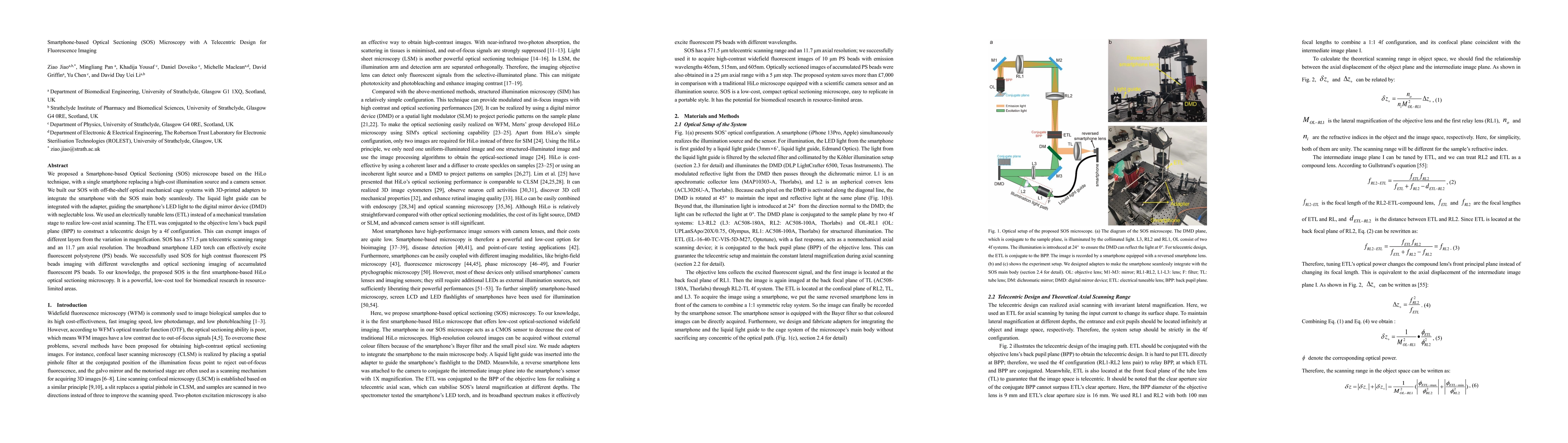
We proposed a Smartphone-based Optical Sectioning (SOS) microscope based on the HiLo technique, with a single smartphone replacing a high-cost illumination source and a camera sensor.We built our SOS with off-the-shelf optical mechanical cage systems with 3D-printed adapters to integrate the smartphone with the SOS main body seamlessly.The liquid light guide can be integrated with the adapter, guiding the smartphone LED light to the digital mirror device with neglectable loss.We used an electrically tunable lens (ETL) instead of a mechanical translation stage to realize low-cost axial scanning. The ETL was conjugated to the objective lens back pupil plane (BPP) to construct a telecentric design by a 4f configuration. This can exempt images of different layers from the variation in magnification. SOS has a 571.5 {\mu}m telecentric scanning range and an 11.7 {\mu}m axial resolution. The broadband smartphone LED torch can effectively excite fluorescent polystyrene (PS) beads. We successfully used SOS for high contrast fluorescent PS beads imaging with different wavelengths and optical sectioning imaging of accumulated fluorescent PS beads. To our knowledge, the proposed SOS is the first smartphone-based HiLo optical sectioning microscopy. It is a powerful, low-cost tool for biomedical research in resource-limited areas.
AI Key Findings
Get AI-generated insights about this paper's methodology, results, significance, and more — seven facets brought into focus.
Impact
Paper Details
Authors
PDF Preview
Key Terms
Citation Network
Current paper (gray), citations (green), references (blue)
Display is limited for performance on very large graphs.
Discussion 0