ArxivLens

Quick Summary:

This study investigates the microstructural features detectable by double diffusion encoding (DDE) MRS in the mouse brain at 11.7T, finding that while traditional infinite cylinder models poorly fit experimental data, incorporating branched fiber structures significantly improves the interpretation of DDE signals. Additionally, short mixing time experiments suggest potential sensitivity to cell body diameter.

arXiv Id: 1908.00317
Date Published: 2019-08-02
Date Updated: 2019-08-02

Categories:

Key Terms (TF-IDF):

cell (0.261)
cell (0.261)
fiber (0.242)
fiber (0.220)
diffusion (0.218)
diameter (0.215)
diffusion (0.208)
diameter (0.207)
signal (0.185)
brain (0.182)
signal (0.178)
angular (0.177)
angular (0.172)
encoding (0.171)
brain (0.169)
encoding (0.162)
mixing (0.150)
influence (0.148)
mixing (0.143)
influence (0.138)
License: http://arxiv.org/licenses/nonexclusive-distrib/1.0/

Summary:

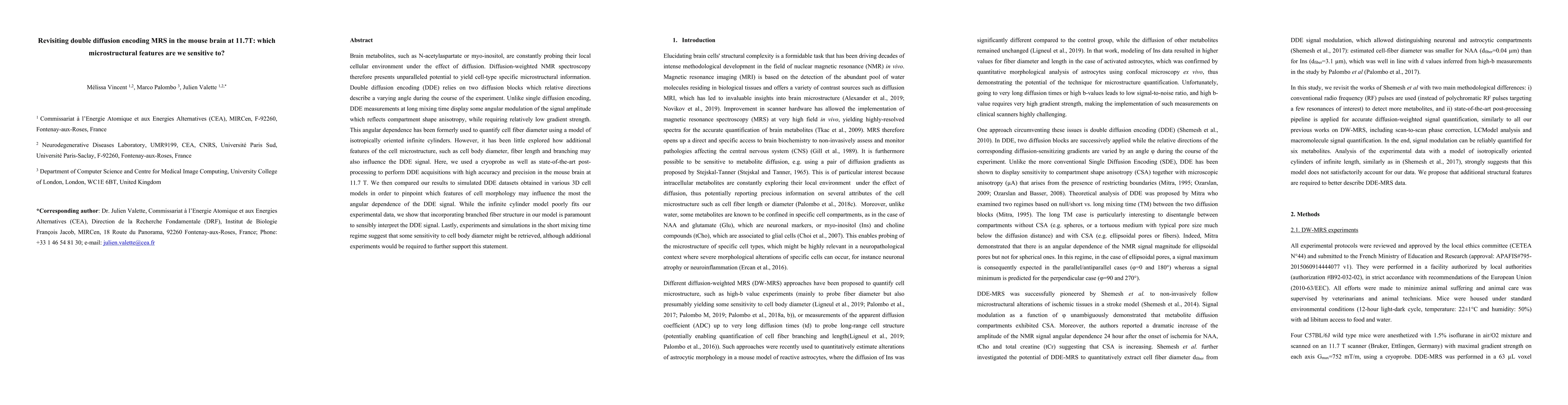
Brain metabolites, such as N-acetylaspartate or myo-inositol, are constantly probing their local cellular environment under the effect of diffusion. Diffusion-weighted NMR spectroscopy therefore presents unparalleled potential to yield cell-type specific microstructural information. Double diffusion encoding (DDE) relies on two diffusion blocks which relative directions describe a varying angle during the course of the experiment. Unlike single diffusion encoding, DDE measurements at long mixing time display some angular modulation of the signal amplitude which reflects compartment shape anisotropy, while requiring relatively low gradient strength. This angular dependence has been formerly used to quantify cell fiber diameter using a model of isotropically oriented infinite cylinders. However, it has been little explored how additional features of the cell microstructure, such as cell body diameter, fiber length and branching may also influence the DDE signal. Here, we used a cryoprobe as well as state-of-the-art post-processing to perform DDE acquisitions with high accuracy and precision in the mouse brain at 11.7 T. We then compared our results to simulated DDE datasets obtained in various 3D cell models in order to pinpoint which features of cell morphology may influence the most the angular dependence of the DDE signal. While the infinite cylinder model poorly fits our experimental data, we show that incorporating branched fiber structure in our model is paramount to sensibly interpret the DDE signal. Lastly, experiments and simulations in the short mixing time regime suggest that some sensitivity to cell body diameter might be retrieved, although additional experiments would be required to further support this statement.

Citation & Reference Network Visualization

This graph shows how this paper is connected to other research. The current paper is shown in dark gray, papers that cite it are in green, and papers it references are in blue. You can:

Current Paper
Papers citing this work (Citations)
Papers cited by this work (References)

• Hover over any node to see the paper title

• Click a node to view that paper's details

• Drag nodes to rearrange the graph

• Use the zoom controls (bottom right) to zoom in/out

Citations and References

Title Authors Year Actions
Introducing µGUIDE for quantitative imaging via generalized uncertainty-driven inference using deep learning
Maëliss Jallais, Marco Palombo
2024
View Paper
Age-trajectories of higher-order diffusion properties of major brain metabolites in cerebral and cerebellar grey matter using in vivo diffusion-weighted MR spectroscopy at 3T
Kadir Şimşek, Cécile Gallea, Guglielmo Genovese, Stéphane Lehéricy, F. Branzoli, Marco Palombo
2024
View Paper
Cerebellar imaging with diffusion magnetic resonance imaging: approaches, challenges, and potential
Henrik Lundell, Christopher J Steele
2024
View Paper
$\mu$GUIDE: a framework for quantitative imaging via generalized uncertainty-driven inference using deep learning
Maëliss Jallais, Marco Palombo
2023
View Paper
Investigating exchange, structural disorder, and restriction in gray matter via water and metabolites diffusivity and kurtosis time-dependence
Eloïse Mougel, J. Valette, M. Palombo
2023
View Paper
Diffusion‐weighted MR spectroscopy: Consensus, recommendations, and resources from acquisition to modeling
Clémence Ligneul, C. Najac, André Döring, C. Beaulieu, F. Branzoli, William T. Clarke, C. Cudalbu, Guglielmo Genovese, S. Jbabdi, I. Jelescu, D. Karampinos, R. Kreis, H. Lundell, M. Marjańska, Harald E Möller, J. Mosso, Eloïse Mougel, S. Posse, S. Ruschke, Kadir Şimşek, Filip Szczepankiewicz, A. Tal, C. Tax, Georg Oeltzschner, M. Palombo, I. Ronen, J. Valette
2023
View Paper
High‐field ex vivo and in vivo two‐dimensional nuclear magnetic resonance spectroscopy in murine brain: Resolving and exploring the molecular environment
Ricardo P. Martinho, Mukul G. Jain, L. Frydman
2022
View Paper
In vivo Correlation Tensor MRI reveals microscopic kurtosis in the human brain on a clinical 3T scanner
Lisa Novello, R. Henriques, A. Ianuş, T. Feiweier, N. Shemesh, J. Jovicich
2021
View Paper
SPHERIOUSLY? The challenges of estimating sphere radius non-invasively in the human brain from diffusion MRI
M. Afzali, M. Nilsson, M. Palombo, Derek K. Jones
2021
View Paper
Characterizing extracellular diffusion properties using diffusion‐weighted MRS of sucrose injected in mouse brain
Mélissa Vincent, Mylène Gaudin, C. Lucas-Torres, A. Wong, C. Escartin, J. Valette
2021
View Paper
Compartmental diffusion and microstructural properties of human brain gray and white matter studied with double diffusion encoding magnetic resonance spectroscopy of metabolites and water
H. Lundell, C. Najac, Marjolein Bulk, H. Kan, A. Webb, I. Ronen
2020
View Paper
SPHERIOUSLY? The challenges of estimating spherical pore size non-invasively in the human brain from diffusion MRI
M. Afzali, M. Nilsson, M. Palombo, Derek K. Jones
2020
View Paper
Double diffusion encoding and applications for biomedical imaging
R. Henriques, M. Palombo, S. Jespersen, N. Shemesh, H. Lundell, A. Ianuş
2020
View Paper
Mapping complex cell morphology in the grey matter with double diffusion encoding MR: A simulation study
A. Ianuş, D. Alexander, Hui Zhang, M. Palombo
2020
View Paper
Challenges for biophysical modeling of microstructure
I. Jelescu, M. Palombo, F. Bagnato, K. Schilling
2020
View Paper
Cytosolic diffusivity and microscopic anisotropy of N‐acetyl aspartate in human white matter with diffusion‐weighted MRS at 7 T
H. Lundell, C. Ingo, T. Dyrby, I. Ronen
2020
View Paper
SANDI: A compartment-based model for non-invasive apparent soma and neurite imaging by diffusion MRI
M. Palombo, A. Ianuş, Michele Guerreri, Daniel Nunes, D. Alexander, N. Shemesh, Hui Zhang
2019
View Paper
μGUIDE: a framework for microstructure imaging via generalized uncertainty-driven inference using deep learning
Maëliss Jallais, Marco Palombo
2023
View Paper

Comments (0)

This website uses cookies to ensure you get the best experience. Learn more