Towards Routine AI-Based PET/CT and SPECT/CT Lesion Segmentation and Tracking in PSMA Theranostics
Publication
Metrics
Paper Preview
Abstract
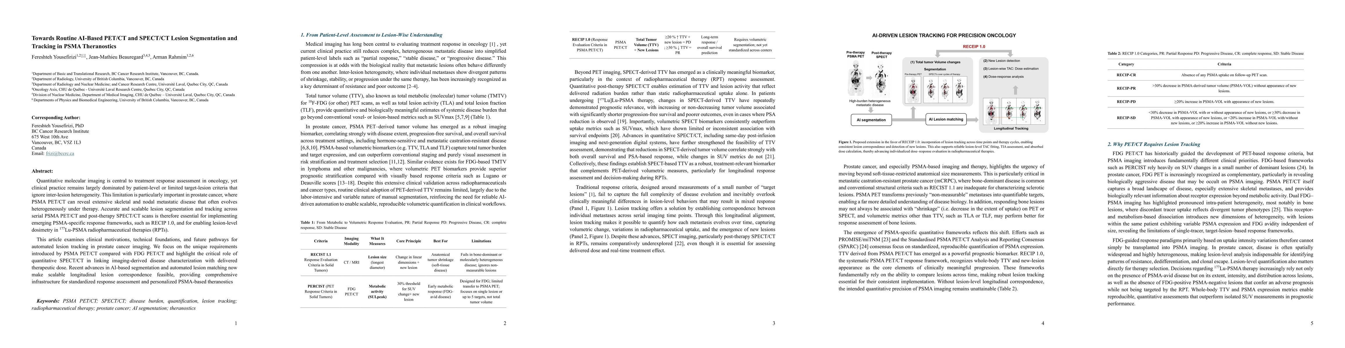
Quantitative molecular imaging is central to treatment response assessment in oncology, yet clinical practice remains largely dominated by patient-level or limited target-lesion criteria that ignore inter-lesion heterogeneity. This limitation is particularly important in prostate cancer, where PSMA PET/CT can reveal extensive skeletal and nodal metastatic disease that often evolves heterogeneously under therapy. Accurate and scalable lesion segmentation and tracking across serial PSMA PET/CT and post-therapy SPECT/CT scans is therefore essential for implementing emerging PSMA-specific response frameworks, such as RECIP 1.0, and for enabling lesion-level dosimetry in 177Lu-PSMA radiopharmaceutical therapies (RPTs). This article examines clinical motivations, technical foundations, and future pathways for automated lesion tracking in prostate cancer imaging. We focus on the unique requirements introduced by PSMA PET/CT compared with FDG PET/CT and highlight the critical role of quantitative SPECT/CT in linking imaging-derived disease characterization with delivered therapeutic dose. Recent advances in AI-based segmentation and automated lesion matching now make scalable longitudinal lesion correspondence feasible, providing comprehensive infrastructure for standardized response assessment and personalized PSMA-based theranostics
AI Key Findings
Get AI-generated insights about this paper's methodology, results, significance, and more — seven facets brought into focus.
Discussion 0