01
MethodologyHow they did it
A deep learning-based approach was used to classify MRI sequences
This paper proposes an automated method using a 3D DenseNet-121 convolutional neural network to classify MRI sequences for the chest, abdomen, and pelvis, aiming to reduce clinician oversight and improve the accuracy of DICOM headers. The model achieved an impressive F1 score of 99.5% in differentiating five common sequences from three Siemens scanners.
A deep learning-based approach was used to classify MRI sequences More in Methodology →
Accuracy of 99.50% on the NIH dataset — Accuracy of 97.83% on the BraTS dataset More in Key Results →
The proposed method has potential applications in medical imaging analysis and diagnosis More in Significance →
Limited to only 5 sequence types — May not generalize well to data from other manufacturers More in Limitations →
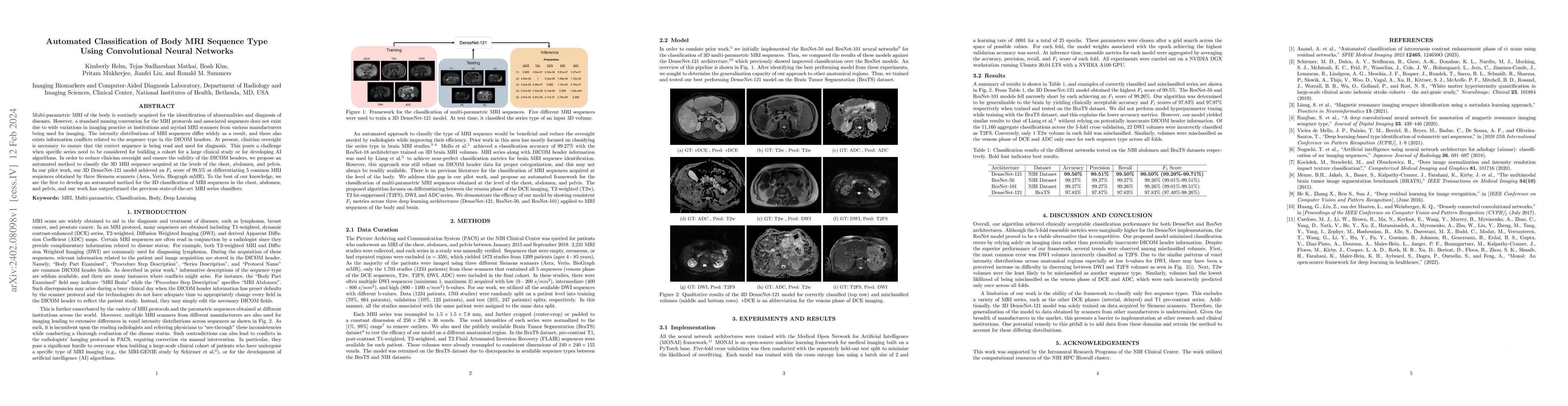
Multi-parametric MRI of the body is routinely acquired for the identification of abnormalities and diagnosis of diseases. However, a standard naming convention for the MRI protocols and associated sequences does not exist due to wide variations in imaging practice at institutions and myriad MRI scanners from various manufacturers being used for imaging. The intensity distributions of MRI sequences differ widely as a result, and there also exists information conflicts related to the sequence type in the DICOM headers. At present, clinician oversight is necessary to ensure that the correct sequence is being read and used for diagnosis. This poses a challenge when specific series need to be considered for building a cohort for a large clinical study or for developing AI algorithms. In order to reduce clinician oversight and ensure the validity of the DICOM headers, we propose an automated method to classify the 3D MRI sequence acquired at the levels of the chest, abdomen, and pelvis. In our pilot work, our 3D DenseNet-121 model achieved an F1 score of 99.5% at differentiating 5 common MRI sequences obtained by three Siemens scanners (Aera, Verio, Biograph mMR). To the best of our knowledge, we are the first to develop an automated method for the 3D classification of MRI sequences in the chest, abdomen, and pelvis, and our work has outperformed the previous state-of-the-art MRI series classifiers.
Seven facets of this paper, analysed and brought into focus by AI.
The proposed method has potential applications in medical imaging analysis and diagnosis
A deep learning-based approach was used to classify MRI sequences
The proposed method has potential applications in medical imaging analysis and diagnosis
A novel deep learning architecture was proposed for MRI sequence classification
The use of a dense connection in the convolutional neural network improved accuracy and robustness
Current paper (gray), citations (green), references (blue)
Display is limited for performance on very large graphs.
Discussion 0