Simulating dark-field x-ray microscopy images with wave front propagation techniques
Publication
Metrics
AI Quick Summary
This paper presents numerical simulations of Dark-Field X-ray Microscopy images using dynamical Takagi-Taupin Equations and wave front propagation techniques. The approach is validated by comparing simulated images to experimental data from a diamond crystal with a stacking fault defect.
Paper Preview
Abstract
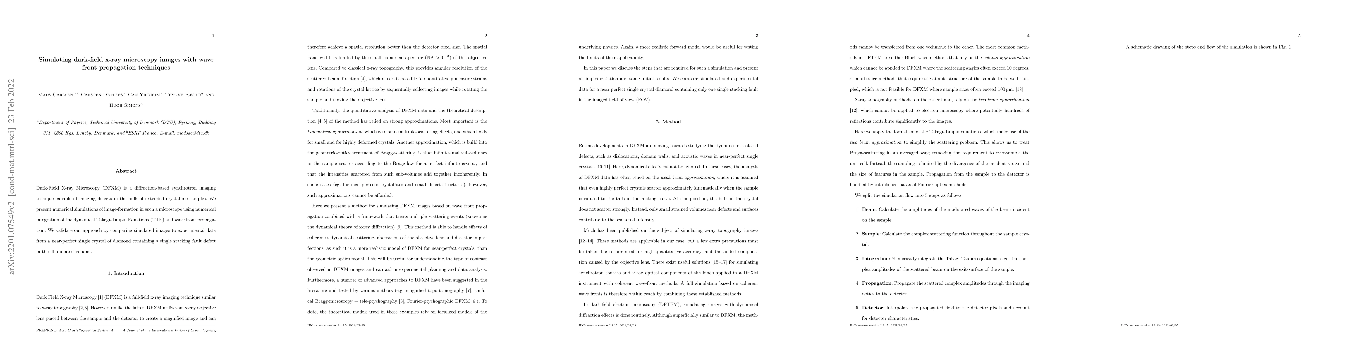
Dark-Field X-ray Microscopy (DFXM) is a diffraction-based synchrotron imaging techique capable of imaging defects in the bulk of extended crystalline samples. We present numerical simulations of image-formation in such a microscope using numerical integration of the dynamical Takagi-Taupin Equations (TTE) and wave front propagation. We validate our approach by comparing simulated images to experimental data from a near-perfect single crystal of diamond containing a single stacking fault defect in the illuminated volume.
AI Key Findings
Get AI-generated insights about this paper's methodology, results, significance, and more — seven facets brought into focus.
Impact
Paper Details
Authors
PDF Preview
Key Terms
Citation Network
Current paper (gray), citations (green), references (blue)
Display is limited for performance on very large graphs.
Discussion 0